
Abstract
Background
The transplantation of exosomes derived from human adipose-derived mesenchymal stem cells (hADSCs) has emerged as a prospective cellular-free therapeutic intervention for the treatment of neurodevelopmental disorders (NDDs), as well as autism spectrum disorder (ASD). Nevertheless, the efficacy of hADSC exosome transplantation for ASD treatment remains to be verified, and the underlying mechanism of action remains unclear.
Results
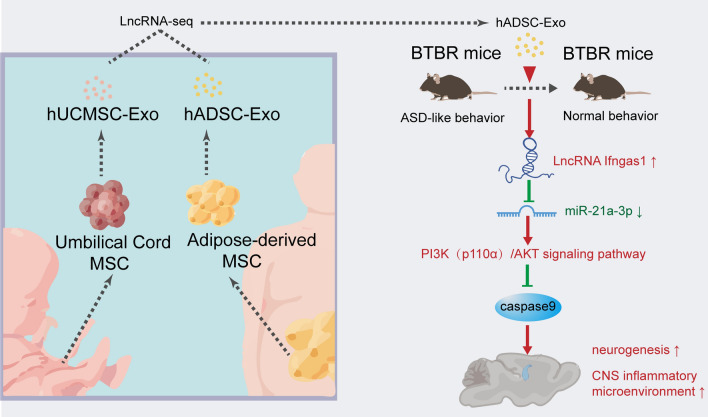
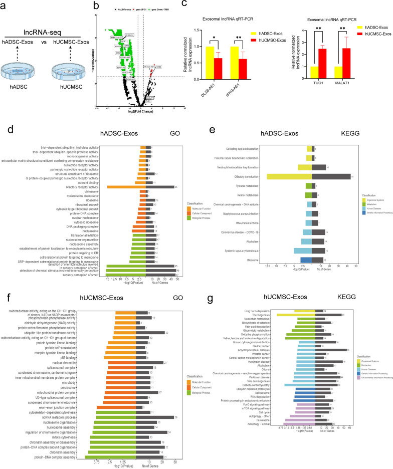
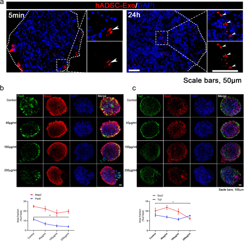
The exosomal long non-coding RNAs (lncRNAs) from hADSC and human umbilical cord mesenchymal stem cells (hUCMSC) were sequenced and 13,915 and 729 lncRNAs were obtained, respectively. The lncRNAs present in hADSC-Exos encompass those found in hUCMSC-Exos and are associated with neurogenesis. The biodistribution of hADSC-Exos in mouse brain ventricles and organoids was tracked, and the cellular uptake of hADSC-Exos was evaluated both in vivo and in vitro. hADSC-Exos promote neurogenesis in brain organoid and ameliorate social deficits in ASD mouse model BTBR T + tf/J (BTBR). Fluorescence in situ hybridization (FISH) confirmed lncRNA Ifngas1 significantly increased in the prefrontal cortex (PFC) of adult mice after hADSC-Exos intraventricular injection. The lncRNA Ifngas1 can act as a molecular sponge for miR-21a-3p to play a regulatory role and promote neurogenesis through the miR-21a-3p/PI3K/AKT axis.
Conclusion
We demonstrated hADSC-Exos have the ability to confer neuroprotection through functional restoration, attenuation of neuroinflammation, inhibition of neuronal apoptosis, and promotion of neurogenesis both in vitro and in vivo. The hADSC-Exos-derived lncRNA IFNG-AS1 acts as a molecular sponge and facilitates neurogenesis via the miR-21a-3p/PI3K/AKT signaling pathway, thereby exerting a regulatory effect. Our findings suggest a potential therapeutic avenue for individuals with ASD.
Introduction
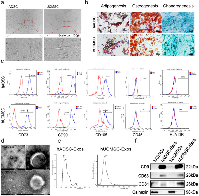
Derived from adipose, umbilical cord and bone marrow tissues and organs, Mesenchymal stem cells (MSCs) are considered a prolific source for tissue engineering and regenerative medicine. Exosomes, which range in size from 30–150 nm and contain a diverse array of proteins, mRNAs, long noncoding RNAs (lncRNAs), and other macromolecules. Two distinct exosome types, hADSC-Exos and hUCMSC-Exos, offer numerous advantages over MSCs, including the retention of parent cell neuroprotection function, long-term stability, minimal immunological rejection, easily internalized into receptor cells, and lower probability of tumor development. Consequently, hADSC-Exos and hUCMSC-Exos have emerged as a prospective cell-free treatment strategy for intervening in brain diseases. Nevertheless, the heterogeneity in hADSC-Exos and hUCMSC-Exos remains unclear.
Exosomes contain lncRNAs, which have been shown to play a key part in regulating neurogenesis in individuals with ASD. Through their ceRNA activity, lncRNAs can act as microRNAs (miRNAs) sponges and thereby contribute to endogenous neurogenesis, apoptosis, neural plasticity, and immune modulation. A majority of ASD brains exhibit a common pattern of lncRNAs dysregulation, such as PTCHD1AS 1-3, SHANK2-AS and BDNF-AS. Exosomes approximately reflect the intracellular status of their host cells, which implies their heterogeneity in different tissue source. However, the role of exosomal lncRNAs is inadequately understood in different tissue source.
The core symptoms of ASD are impaired social interaction, impaired communication, and repetitive stereotyped behavior disorder, a heterogeneous developmental disorder. The potential neurobiological etiology of ASD includes GABAergic imbalances, impaired neurogenesis and neuroimmune processes. The prevalence of ASD is increasing year by year, but currently, there is no established standard drug for patients with ASD. Stem cell-based regenerative therapy has been widely concerned for their ability to treat diverse range of neurological disorders. Recent studies have shown that transplantation of hematopoietic stem cells (HSCs) from the fetal liver is beneficial in alleviating ASD-like symptoms in children. Transplantation of human amniotic epithelial cells (hAECs) corrects social deficits in BTBR mice, the specific mechanism of which is related to the promotion of hippocampal neurogenesis. Despite the disclosure of the advantageous impact of MSC on the fundamental symptoms of BTBR mice, the precise mechanism through which MSC-Exo confers benefits to neurogenesis remains undisclosed.
In the present study, the lncRNA-seq of hADSC-Exos and hUCMSC-Exos to elucidate the functional diversity of MSC-Exos. Subsequently, the underlying mechanism responsible for the neuroprotective properties of hADSC-Exos was explored. In vitro experiments demonstrated that hADSC-Exos substantially enhanced the accumulation of neural progenitor cells (NPCs) and promoted neuron survival in brain organoids. In vivo, hADSC-Exos mitigated stereotyped and anxiety behavior, impaired new object recognition, and social deficits in BTBR mice. The administration of hADSC-Exos demonstrated the ability to ameliorate neurodevelopmental abnormalities and suppress the inflammatory microenvironment within the brains of BTBR mice. MiR-21a-3p expression was markedly upregulated in BTBR mice based on qRT-PCR results, which was effectively ameliorated by the intervention with hADSC-Exos. These findings indicate a potential regulator role of hADSC-Exos in the process of neurogenesis.
Results
Characterization of exosomes derived from hADSC and hUCMSC
LncRNAs sequencing analysis to identify hADSC-Exo and hUCMSC-Exo
hADSC-Exo suppress proliferation of NSC and tend to promote neurogenesis in brain organoids
hADSC-Exo can regulate the expression of neurogenesis and brain inflammation-related genes in BTBR mice
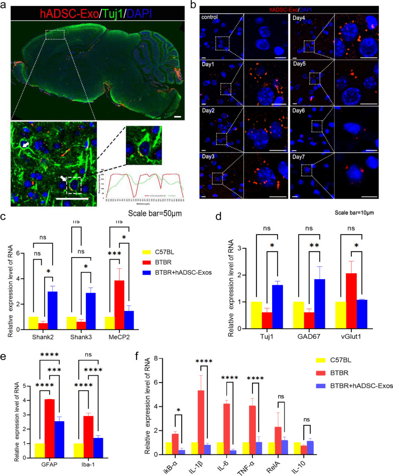
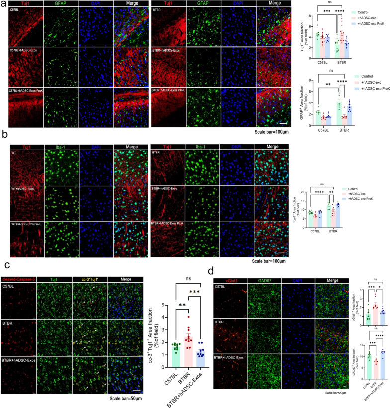
hADSC-Exo treated ameliorate inflammation and promote neurogenesis in PFC regions of BTBR mice brain
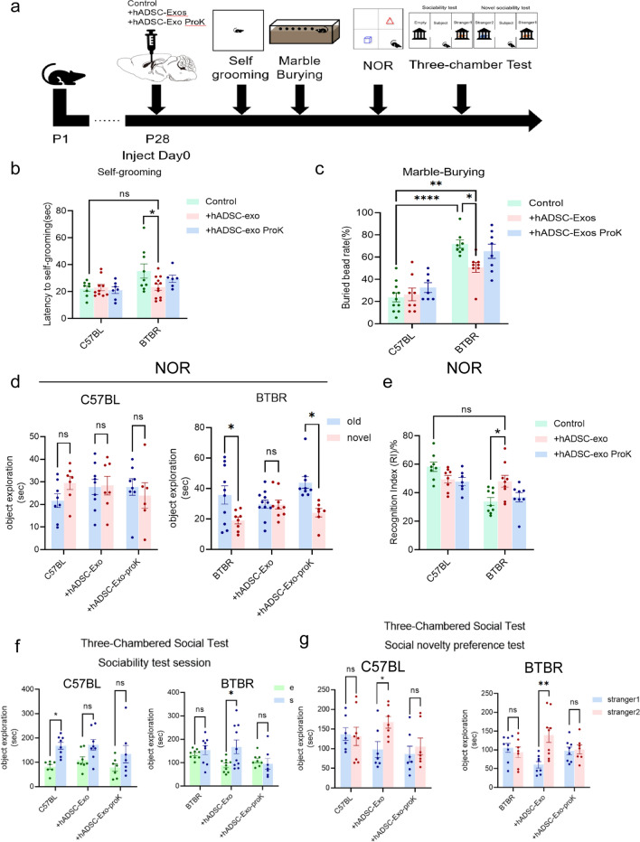
hADSC-Exo treated reverses ASD-like behavior in BTBR mice
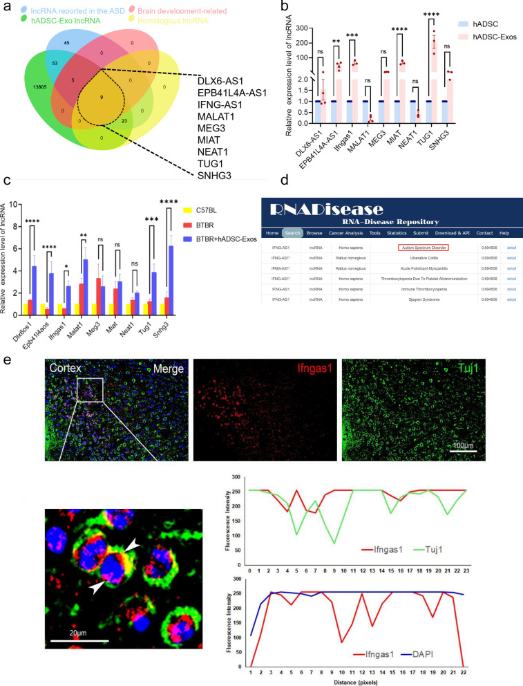
The lncRNA IFNGAS1 has been identified as significantly associated with ASD in hADSC-Exo
Ifngas1 may act as a molecular sponge of miR-21a-3p, thereby negatively regulating Akt signaling pathway
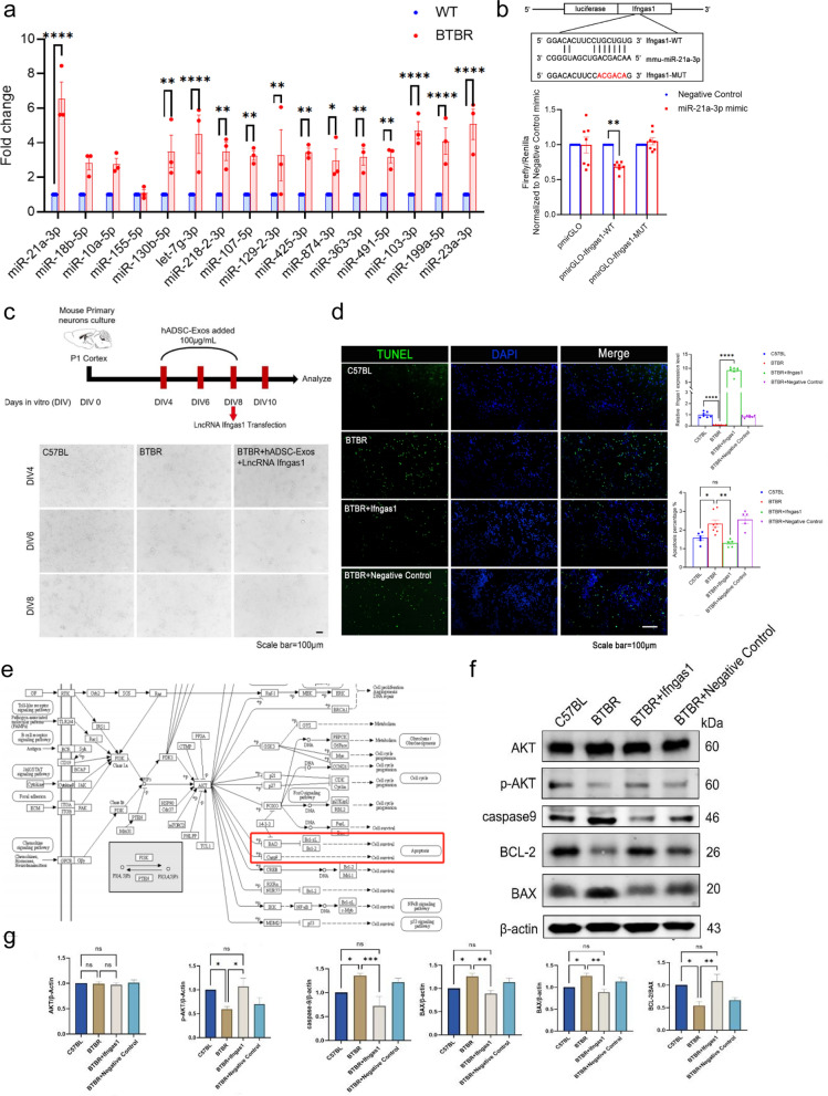
Discussion
Compared with other conventional synthesized drug, exosomes have emerged as a promising therapeutic option for neurological diseases due to their ability to cross the blood–brain barrier. For cellular therapy products to be safe and effective, comprehensive characterization, identification of the most relevant critical quality attributes (CQAs), and quality control are necessary. Researchers have devoted substantial efforts to elucidate the biological properties of exosomes and their components and their roles in central nervous system disease, such as AD, Parkinson's disease (PD) and Huntington's disease (HD). At the same time, several exosome databases have been created. ExoRBase 2.0 database have shown that exosomes in the human biofluids also contain different lncRNA. LncExpDB documents exosomal lncRNAs differentially expressed across diverse biological conditions. EVAtlas houses the expression profiles of ncRNA types in EV samples from human tissues, but not yet for lncRNAs. Exosomes may carry specific lncRNAs and miRNAs from a variety of tissue sources. Understanding the origin of exosomes is essential to gain insight into the prospective applications of human exosomes in neurotherapeutics. However, intervention studies to date on the ASD have not fully paid attention to the origin of exosomes. Here, the two types of widely clinical used hMSCs and their exosome were obtained, and the lncRNA-seq of hADSC-Exo and hUCMSC-Exo was performed. Due to the key words “Olfactory transduction”, “Alcoholism” and “Staphylococcus aureus infection” in hADSC-Exo lncRNA-related KEGG analysis, we were reminded of its nervous-immune regulation ability (Fig. 2e). Hence, hADSC-Exo was selected to continue the study. Notably, there were only two biological replicates in hADSC-Exo. One hADSC-Exo 2 sample was excluded from the analysis since it might be contaminated. The Veen analysis shows similar patterns for hADSC-Exo1 and 3 samples (Additional file 1: Fig. S2c and d). LncRNAs intersect in three hUCMSC-Exo samples only 67 lncRNAs (Additional file 1: Fig. S2e). We used the union to do the comparison showed all differentially expressed lncRNA detected in hADSC-Exo and hUCMSC-Exo (Additional file 1: Fig. S2f). This prompts future research with larger samples of MSC-Exo, as the main limitation of our study is the low number of included hADSC-Exo and hUCMSC-Exo biological duplication. Verification of sequencing data through qRT-PCR is a key step in lncRNA-seq analyses. Among them, some lncRNAs were randomly selected for qRT-PCR, and the qRT-PCR results were consistent with the sequencing data, such as DLX6-AS1 and IFNG-AS1 (Fig. 2c).
Recently, Samir EL Andaloussi et al. investigated the effects of different exosome sources and doses on recipient cells in a systematic manner. They found that a low dose of exosomes produces profound transcriptional changes specific to the exosome cell source, while a high dose of exosomes produces a standardized response. High doses would likely overload the endocytic machinery and do not represent physiological conditions. Low doses would contain the fewest exosome-derived transcripts may not be able to be treated. Their study highlights that standardization and comparable dosages should become common practice in exosome studies. Here, an analysis of bioinformatics-based cell sources was conducted, and doses were selected based on human brain organoids. Hence, we decided to choose an intermediate concentration. Our current study provides support for the standardization and comparable dosages of exosome paradigm to conducting initial clinical research.
The ASD mouse model BTBR mouse model recapitulates many features of the human ASD phenotype, such as GABAergic imbalances. Functional analyses show that the genes differentially expressed in the cerebral cortex of BTBR mice are mainly involved in the following biological processes: "neurological development", "social behavior", etc.. The results of this analysis further demonstrated the similarity between the BTBR mouse model and ASD patients. Notably, in this study, we innovatively explored the efficient of hADSC-Exo in human brain organoid. Findings from human brain organoid and ASD mouse model provide the foundation for subsequent investigations in more complex systems. Due to the lack of approved drugs to treat the symptoms of ASD, hADSC-Exo might be a good therapeutic agent in future.
It remains unclear how MSC-Exo contributes to neurogenesis in ASD, despite previous studies investigating its role in the disorder. Elucidation of the mechanisms by which exosomal lncRNA ameliorate ASD-like behavior and neurogenesis is essential for intervention effectiveness. According to Joerger-Messerli et al., EVs derived from human Wharton's jelly MSC (hWJ-MSC)-MSCs may prevent and resolve HI-induced apoptosis in neurons in the neonatal brain by transferring let-7-5p from the EVs. In this study, we illustrated that hADSC-Exos activated the PI3K(p110α)/AKT signaling pathway through the Ifngas1/miR-21a-3p axis. It can ameliorate neurogenesis and ASD-like behavior in BTBR mice. Exosomes contain lncRNAs and many other macromolecules from their source cells, such as proteins, miRNAs, etc. Further investigation of hADSC-Exos-mediated neuroprotection and neurogenesis mechanisms will be needed before considering ASD-related clinical practice. Moreover, it may be worth to test MSC-exo effect in ASD by other high-throughput approaches, including LC–MS proteome analysis and single cell sequencing as well. Overall, our findings add further insights into the hADSC-Exo functions in ASD.
Materials and methods
Ethics statements
All procedures followed the guidelines of the National Health and Medical Research Council of China and received approval from the Animal Ethics Review Committee of Tongji University.
Cell culture
A healthy donor provided written informed consent to the East Hospital Affiliated to Tongji University to obtain the human adipose tissue samples. The ADSC cells in this study were utilized and the cells were serviced under the same conditions as those described in our previous study. Human umbilical cord tissue samples were acquired from the Stem Cell Bank of the East Hospital Affiliated to Tongji University. After obtaining informed consent from the mother and her family, the donor signed a written informed statement. The approval was obtained from the East Hospital Ethical Review Board. All cells were incubated at 37 °C in an incubator with 5% CO2.
Exo isolation and characterization
Exosomes isolated from the cell supernatant of hADSC/hUCMSC in the same methodology as described previously. The cells were separated from the culture medium by centrifugation at 300g for 10 min. Transfer the supernatant to the new centrifugal tube and centrifuge for 10 min at 2000g, followed by an additional 30 min at 10,000g. The liquid supernatant was then centrifuged at 1,000,000g for 2 h at 4 °C (Additional file 1: Fig. S1). Afterward, exosomes were suspended in 1 × PBS and kept at − 80 °C for subsequent experiments.
Exo characterization
The protein in exosome was measured to quantify exosomes. The concentration of Exo was evaluated using the bicinchoninic acid (BCA, Thermo Fisher Scientific, Waltham, MA, USA). In Fig. 1f, hADSCs and hUCMSCs samples of whole-cell lysate (WCL) was reserved. The WCL were boiled with SDS–PAGE sample loading buffer, separated by SDS–PAGE, blotted on PVDF membranes. Western Blot with anti-CD9, anti-63 and anti-CD81 is used for identifying exosome. Transmission electron microscopy (TEM, FEI, USA) was used to examine the morphology of the isolated exosomes. Nanosight tracking analysis (NTA, Malvern, USA) was performed to analyze exosome morphology. In order to analyze particle numbers, the Nanoparticle Tracking Analysis (NTA) 3.0 software was used. hADSC-Exo was labeled with PKH26 using the PKH26 Red Fluorescent Cell Linker Mini Kit (Sigma, St Louis, MO, USA). Proteinase K used for chemical disruption of hADSC-Exo (Sigma, St Louis, MO, USA).
Exo lncRNA-seq
The total RNAs were enriched from exosomes of hADSC and hUCMSC were extracted by RiboBio Co., Ltd, Guangzhou, China. Six RNA samples (hADSC-Exo and hUCMSC-Exo) were used in the subsequent analysis. Contaminated sequencing batch of hADSC-Exo2 samples were excluded. Normalized expression levels of the genes between the hADSC-Exo1 and 3 are well correlated with the Pearson correlation coefficient (R) values more than 0.99 (Additional file 1: Fig. S2b and c). In this paper, the differentially expressed lncRNAs were determined by |log2(FoldChange)|> 1 and Qvalue < 0.05, with thresholds for up- and down-regulated lncRNAs.
Generation of cerebral organoids
The H9 human embryonic stem cell (H9 ES cell) used in this study was were donated by Professor Ru Zhang from Tongji University. H9 ES cells were cultured on Vitronectin XF™ (stem cell technologies, Canada) with mTeSR™ medium (stem cell technologies, Canada). After getting enough high-quality and low-differentiated H9 ES cells, then using the STEMdiff Cerebral Organoid kit (stem cell technologies, Canada). Briefly, on day 0, H9 ES cells were dissociated into single cells with ACCUTASE™ (stem cell technologies, Canada) and then suspended in embryoid body (EB) Seeding Medium. Every EB was embedded in 15 µL of Cultrex UltiMatrix (R&D SYSTEMS, USA) and transferred into 6-well ultra-low attachment plate containing 3 mL expansion medium. After 10 d, embedded organoids' culture solution was replaced with maturation medium and organoids were placed on orbital shaker in 37℃ incubators at the speed of 65 rpm until day 50. For exosome co-cultured, based on the reference dose in literature, a concentration gradient is established to determine the optimal concentration.
Mice
Adult pairs of BTBR mice were acquired from The Jackson Laboratory (Bar Harbor, Maine) and were subsequently bred. The mice were housed in an SPF animal facility, where they were kept in white plastic enclosures with unlimited access to water and food.
Exo transplantation
At the age of 4 weeks, BTBR male mice were fixed in a stereotactic frame (Ruiwode, Shenzhen, Guangdong Province, China). In the presence of 4% isoflurane, hADSC-exo, 2 μl per injection site, were injected into the cerebral lateral ventricles bilaterally at 0.5 μL/min (Hamilton 701N syringe) to the following coordinates (relative to the bregma): anterior–posterior, − 1 mm; medial–lateral, ± 0.8 mm; dorsal–ventral, − 1.5 mm. Ten minutes after the needle was inserted, it was withdrawn. Animals were also treated with 0.3% gentamicin for 3 days around transplantation in order to suppress any possible immune response. The behavioral experiment was done 2 weeks after the last treatment.
Behavioral studies
Repetitive behavior was analyzed by a self-grooming test, in short, the mouse were kept in an empty cage (30 × 30 × 29.5 cm) for 10 min, during which time their self-grooming behavior was recorded.
Anxiety-like behavior was analyzed by marble-burying test, in short, twenty clean marbles (d = 14 mm) were evenly distributed on the surface of the corncob cushion (29 × 18 × 13.5 cm) at a depth of 5 cm. After 30 min of acclimatization in the test room, the mice were then placed in the test cage containing the marbles. The marbles were counted after 30 min of exploration.
The New Object Recognition test was executed in the opaque walled box (30 × 30 × 29.5 cm). The mouse was given 10 min to explore the arena without any objects before the adaptive test. Then, two familiar objects were fixed in the box and the mouse could explore two familiar objects for 10 min during the adaptive session. In the test session, a familiar object was changed by a new one, the mouse could explore all the objects for 10 min. The time spent by the mice exploring the novel object was analyzed and recorded. Recognition index = novel object recognition time/total object recognition time.
As previously described, the three-chamber test was employed to measure sociability behaviors. In essence, the apparatus comprised three interconnected chambers with entryways. The mice explored the three-chambered apparatus for 10 min as part of the adaptation task. Afterward, the mice were allowed to interact either with an empty wire mesh cylinder or an unfamiliar mice#1 called stranger1. Mice were then able to explore the three rooms freely for 10 min. Following that, another mouse, unfamiliar mice#2 were placed in a opposite chamber, which was empty in the previous session, for the social novelty preference test. Finally, the time spent in the different chamber was counted.
Immunohistochemistry (IHC)
Histological examinations were conducted on three mice from each group at random, IHC assay was performed as previously described. Anti-ki67 (1:200, Abcam, UK, ab16667), anti-GFAP (1:1000, Abcam, UK, ab7260), anti-cleaved-caspase-3 antibody (1:500, Servicebio, GB11532), anti-Tuj1 (1:1000, Abcam, UK, ab7751) anti-Nestin (1:2000, Abcam, UK, ab221660), anti-vGlut1 (1:100, Biolegend, MMS-5245), anti-Iba-1 (1:500, Servicebio, GB11105), anti-GAD67 (1:500, Abcam, UK, ab13508). The aforementioned primary antibodies and secondary antibodies were used.
Histology
After BTBR mice were euthanized, the hearts, lungs, kidneys, testes, brains, livers and spleens were dissected. The HE staining was carried out on tissue samples from at least three mice of each genotype. Nissl staining was performed by incubating slides with 0.1% Nissl dye for 10 min. To quantify hippocampal neurons, Golgi-Cox staining was used. The slides were first immersed in 1:1 hydrochloric acid and 100% ethanol for 2 h, then washed under running water.
Dual-luciferase reporter gene assay
To generate the miR-21a-3p target site-containing 3′-UTR sequence of lncRNA Ifngas1, the overexpressing plasmid was used. A 1.5% agarose gel was used to purify the DNA fragments. Using XbaI enzyme-digested vectors pGL3-Control (Promega, Madison, WI, USA) to insert downstream of the luciferase gene. The 293T cells, at 80–90% confluence, were co-transfected with lncRNA Ifngas1 3′-UTR and miR-21a-3p mimic. In vitro transfection was carried out with Xfect Transfection Reagent (Takara Bio, USA). In vivo transfection was performed using in vivo-jetPEI® reagent (Polyplus-transfection SA, France).
Real-time PCR (qRT-PCR)
| Gene name | Gene type | Experiment | Primer (5′-3′) |
|---|---|---|---|
| Gapdh | Total RNA | qRT-PCR Forward | GCTGTCAACGATACGCTACGTAACG |
| qRT-PCR Reverse | TGAAGGGGTCGTTGATCG | ||
| Map2 | Total RNA | qRT-PCR Forward | CAATCTTCACATTACCACCTCCA |
| qRT-PCR Reverse | CTCTAAAGAACATCCGTCAC | ||
| MeCP2 | Total RNA | qRT-PCR Forward | TTCTATTCTGGGCTTTTGATTTGT |
| qRT-PCR Reverse | CCCTTGTCCTACTCTATGGTTATCA | ||
| GFAP | Total RNA | qRT-PCR Forward | CGGAGACGCATCACCTCTG |
| qRT-PCR Reverse | AGGGAGTGGAGGAGTCATTCG | ||
| Tuj1 | Total RNA | qRT-PCR Forward | CAGCGATGAGCACGGCATAGAC |
| qRT-PCR Reverse | CCAGGTTCCAAGTCCACCAGAATG | ||
| Iba-1 | Total RNA | qRT-PCR Forward | ATCCCAAGTACAGCAGTGATGAGG |
| qRT-PCR Reverse | AAATAGCTTTCTTGGCTGGGGGAC | ||
| Syn1 | Total RNA | qRT-PCR Forward | CATTCTGGGATGGGCAAGGTCAAG |
| qRT-PCR Reverse | GGCTCAGCAGTGGCATATGTCTTAG | ||
| GAD67 | Total RNA | qRT-PCR Forward | TGCGCTTGGCTTTGGAA |
| qRT-PCR Reverse | TCCCCCTTTCATTGCACTTT | ||
| vGlut1 | Total RNA | qRT-PCR Forward | CTATGTCTAGCAGCTTCG |
| qRT-PCR Reverse | TCAATGTATTTGCTCCT | ||
| Olig2 | Total RNA | qRT-PCR Forward | CGGTGGCTTCAAGTCATCTTCCTC |
| qRT-PCR Reverse | GGGCTCAGTCATCTGCTTCTTGTC | ||
| Shank2 | Total RNA | qRT-PCR Forward | GAGCAGCCACCGTGATGATGAC |
| qRT-PCR Reverse | ACCACCGTCTTGTCCTCAATAATGC | ||
| Shank3 | Total RNA | qRT-PCR Forward | GATGTGCAAACCCGAGACTCTGAG |
| qRT-PCR Reverse | TCCTCTGGTGACTTCCGCTCTTC | ||
| miR-21a-3p | miRNA | qRT-PCR Forward | CAACAGCAGTCGATGGGCT |
| miR-18b-5p | miRNA | qRT-PCR Forward | TAAGGTGCATCTAGTGCTGTTAG |
| miR-10a-5p | miRNA | qRT-PCR Forward | TACCCTGTAGATCCGAATTTGTG |
| miR-155-5p | miRNA | qRT-PCR Forward | TTAATGCTAATTGTGATAGGGGT |
| miR-130b-5p | miRNA | qRT-PCR Forward | ACTCTTTCCCTGTTGCACTACT |
| let-7g-3p | miRNA | qRT-PCR Forward | ACTGTACAGGCCACTGCCTTGC |
| miR-218-2-3p | miRNA | qRT-PCR Forward | CATGGTTCTGTCAAGCACCGCG |
| miR-874-3p | miRNA | qRT-PCR Forward | CTGCCCTGGCCCGAGGGACCGA |
| miR-107-5p | miRNA | qRT-PCR Forward | AGCTTCTTTACAGTGTTGCCTTG |
| miR-129-2-3p | miRNA | qRT-PCR Forward | AAGCCCTTACCCCAAAAAGCAT |
| miR-425-3p | miRNA | qRT-PCR Forward | ATCGGGAATGTCGTGTCCGCC |
| miR-23a-3p | miRNA | qRT-PCR Forward | ATCACATTGCCAGGGATTTCC |
| miR-363-3p | miRNA | qRT-PCR Forward | AATTGCACGGTATCCATCTGTA |
| miR-491-5p | miRNA | qRT-PCR Forward | AGTGGGGAACCCTTCCATGAGG |
| miR-103-3p | miRNA | qRT-PCR Forward | AGCAGCATTGTACAGGGCTATGA |
| miR-199a-5p | miRNA | qRT-PCR Forward | CCCAGTGTTCAGACTACCTGTTC |
| U6 | miRNA | qRT-PCR Forward | CGCTTCGGCAGCACATATAC |
| U6 | miRNA | qRT-PCR Reverse | AATTTGCGTGTCATCCTTGC |
| mmu-Malat1 | LncRNA | qRT-PCR Forward | GCGAGCAGGCATTGTGGAGAG |
| qRT-PCR Reverse | GCCGACCTCAAGGAATGTTACCG | ||
| mmu-Dlx6os1 | LncRNA | qRT-PCR Forward | CTGAAGACTGACTGAGCGTGGAAG |
| qRT-PCR Reverse | TCTGGGCGTAGGTTTCTCTCTGG | ||
| mmu-Ifngas1 | LncRNA | qRT-PCR Forward | GGAGGCTAGTGTCTGGATGTTGTTG |
| qRT-PCR Reverse | AGACTGGTGGCTGCTCTGAACTC | ||
| mmu-Miat | LncRNA | qRT-PCR Forward | TTTGCCTTTCTGGTCTGTTCCTTCC |
| qRT-PCR Reverse | CCGCCATCATCCAAGCCGTTAG | ||
| mmu-Snhg3 | LncRNA | qRT-PCR Forward | TCGCTCTCTTGGTGTGCTTGTTC |
| qRT-PCR Reverse | CCCCGCTGATTCTCTTCTTTCCTC | ||
| mmu-Epb41l4aos | LncRNA | qRT-PCR Forward | GGGCGGGAATAAAGCGAAGACC |
| qRT-PCR Reverse | GGACACCCTTTAACCCACTCTGTG | ||
| mmu-TUG1 | LncRNA | qRT-PCR Forward | TTCCTACCACCTTACTACTGACG |
| qRT-PCR Reverse | GGAGGTAAAGGCCACATC | ||
| mmu-MEG3 | LncRNA | qRT-PCR Forward | TTGCAAGGGGCAAGGACTCTC |
| qRT-PCR Reverse | TCATGTCGTCGGTTGGAAAGG | ||
| mmu-NEAT1 | LncRNA | qRT-PCR Forward | GTTCCGTGCTTCCTCTTCTG |
| qRT-PCR Reverse | CAGGGTGTCCTCCACCTTTA | ||
| hsa-MALAT1 | LncRNA | qRT-PCR Forward | CCAGGTGCTACACAGAAGTGGAT |
| qRT-PCR Reverse | GCTTGCTCGCTTGCTCCTCAG | ||
| hsa-DLX6-AS1 | LncRNA | qRT-PCR Forward | TTCAAGCGATTCTCCTGCCTCAAG |
| qRT-PCR Reverse | CAATGTGGCGAAACCCCGTCTC | ||
| hsa-IFNG-AS1 | LncRNA | qRT-PCR Forward | AAGCCCACCAACTGCTAACAACC |
| qRT-PCR Reverse | ACCCTTCAAAGACTTTCCCAACTGG | ||
| hsa-MIAT | LncRNA | qRT-PCR Forward | ACTAACTCCTGCCTTCCTGGTCTG |
| qRT-PCR Reverse | CCAGCCATGCCGACATCCAAG | ||
| hsa-SNHG3 | LncRNA | qRT-PCR Forward | TGCCTCAGCCTCCCAAGTAGC |
| qRT-PCR Reverse | TGGGCGGATCACGAGGTCAG | ||
| hsa-EPB41L4A-AS1 | LncRNA | qRT-PCR Forward | TCGGTCCCTCACTGGCACTTC |
| qRT-PCR Reverse | CAGGCTTCCGTCCCACAATGC | ||
| hsa-NEAT1 | LncRNA | qRT-PCR Forward | CCAGTGTGAGTCCTAGCATTGC |
| qRT-PCR Reverse | CCTGGAAACAGAACATTGGAGAAC | ||
| hsa-TUG1 | LncRNA | qRT-PCR Forward | ACCGGAGGAGCCATCTTGTC |
| qRT-PCR Reverse | GAAAGAGCCGCCAACCGATC | ||
| hsa-MEG3 | LncRNA | qRT-PCR Forward | TGGCATAGAGGAGGTGAT |
| qRT-PCR Reverse | GGAGTGCTGTTGGAGAATA |
Western blot (WB)
WB was conducted following the methodology described in a previous publication. The following antibodies were applied: Rabbit Anti-CD9 antibody (1:1000, Abcam, UK, ab92726), Rabbit Anti-CD63 antibody (1:1000, Abcam, UK, ab134045), Rabbit Anti-CD81 antibody (1:1000, Abcam, UK, ab109201), Rabbit Anti-Calnexin antibody (1:1000, Abcam, UK, ab22595), Rabbit Anti-Caspase-9 antibody (1:1000, Abcam, UK, ab22595), Rabbit Anti-Phospho-AKT(Ser473) antibody (1:1000, Absin, abs130002), Rabbit Anti-AKT antibody (1:1000, CST, Beverly, MA, USA, 4685S), Rabbit Anti-BAX antibody (1:8000, Proteintech, 50599-2-Ig), Mouse Anti-Bcl2 antibody (1:2000, Proteintech, 68103-1-Ig), Mouse Anti-GAPDH antibody (1:1000, Servicebio, GB15002). Using Odyssey Infrared Imaging System, the original images of the membranes were recorded and analyzed according to the ECL WB Protocol (Bio-Rad, Milan, Italy).
Data analysis
The statistical analysis was performed using GraphPad Prism. In the figure legends, data were presented as the mean ± standard deviation (S.D.) or ± standard error of the mean (S.E.M.). A p value < 0.05 was considered statistically significant. Adobe Illustrator CC software and Figdraw (https://www.figdraw.com/static/index.html) were used to draw the chart for the specific mechanism of hADSC-Exo treatment of ASD.
Sources
- Mizuno M, Endo K, Katano H, Amano N, Nomura M, Hasegawa Y, Ozeki N, Koga H, Takasu N, Ohara OTransplantation of human autologous synovial mesenchymal stem cells with trisomy 7 into the knee joint and 5 years of follow-upStem Cells Transl Med20211015301543 https://doi.org/10.1002/sctm.20-0491
- Zhang Y, Huang X, Sun T, Shi L, Liu B, Hong Y, Fu QL, Zhang Y, Li XMicroRNA-19b-3p dysfunction of mesenchymal stem cell-derived exosomes from patients with abdominal aortic aneurysm impairs therapeutic efficacyJ Nanobiotechnol202321135 https://doi.org/10.1186/s12951-023-01894-3
- Zhu W, Sun L, Zhao P, Liu Y, Zhang J, Zhang Y, Hong Y, Zhu Y, Lu Y, Zhao WMacrophage migration inhibitory factor facilitates the therapeutic efficacy of mesenchymal stem cells derived exosomes in acute myocardial infarction through upregulating miR-133a-3pJ Nanobiotechnol20211961 https://doi.org/10.1186/s12951-021-00808-5
- Yari H, Mikhailova MV, Mardasi M, Jafarzadehgharehziaaddin M, Shahrokh S, Thangavelu L, Ahmadi H, Shomali N, Yaghoubi Y, Zamani MEmerging role of mesenchymal stromal cells (MSCs)-derived exosome in neurodegeneration-associated conditions: a groundbreaking cell-free approachStem Cell Res Ther202213423 https://doi.org/10.1186/s13287-022-03122-5
- Perets N, Oron O, Herman S, Elliott E, Offen DExosomes derived from mesenchymal stem cells improved core symptoms of genetically modified mouse model of autism Shank3BMol Autism20201165 https://doi.org/10.1186/s13229-020-00366-x
- Wu YE, Parikshak NN, Belgard TG, Geschwind DHGenome-wide, integrative analysis implicates microRNA dysregulation in autism spectrum disorderNat Neurosci20161914631476 https://doi.org/10.1038/nn.4373
- Nguyen LS, Fregeac J, Bole-Feysot C, Cagnard N, Iyer A, Anink J, Aronica E, Alibeu O, Nitschke P, Colleaux LRole of miR-146a in neural stem cell differentiation and neural lineage determination: relevance for neurodevelopmental disordersMol Autism2018938 https://doi.org/10.1186/s13229-018-0219-3
- Lu J, Zhu Y, Williams S, Watts M, Tonta MA, Coleman HA, Parkington HC, Claudianos CAutism-associated miR-873 regulates ARID1B, SHANK3 and NRXN2 involved in neurodevelopmentTransl Psychiatry202010418 https://doi.org/10.1038/s41398-020-01106-8
- Hosseini E, Bagheri-Hosseinabadi Z, De Toma I, Jafarisani M, Sadeghi IThe importance of long non-coding RNAs in neuropsychiatric disordersMol Aspects Med201970127140 https://doi.org/10.1016/j.mam.2019.07.004
- de Lima TA, Zuanetti PA, Nunes MEN, Hamad APADifferential diagnosis between autism spectrum disorder and other developmental disorders with emphasis on the preschool periodWorld J Pediatr202319715726 https://doi.org/10.1007/s12519-022-00629-y
- Qin L, Williams JB, Tan T, Liu T, Cao Q, Ma K, Yan ZDeficiency of autism risk factor ASH1L in prefrontal cortex induces epigenetic aberrations and seizuresNat Commun2021126589 https://doi.org/10.1038/s41467-021-26972-8
- Arranz MJ, Salazar J, Bote V, Artigas-Baleri A, Serra-LLovich A, Triviño E, Roige J, Lombardia C, Cancino M, Hernandez MPharmacogenetic interventions improve the clinical outcome of treatment-resistant autistic spectrum disorder sufferersPharmaceutics202214999 https://doi.org/10.3390/pharmaceutics14050999
- Fan Y, Chen Z, Zhang MRole of exosomes in the pathogenesis, diagnosis, and treatment of central nervous system diseasesJ Transl Med202220291 https://doi.org/10.1186/s12967-022-03493-6
- Pistollato F, Forbes-Hernández TY, Calderón Iglesias R, Ruiz R, Elexpuru Zabaleta M, Cianciosi D, Giampieri F, Battino MPharmacological, non-pharmacological and stem cell therapies for the management of autism spectrum disorders: a focus on human studiesPharmacol Res2020152104579 https://doi.org/10.1016/j.phrs.2019.104579
- Zhang R, Cai Y, Xiao R, Zhong H, Li X, Guo L, Xu H, Fan XHuman amniotic epithelial cell transplantation promotes neurogenesis and ameliorates social deficits in BTBR miceStem Cell Res Ther2019101153 https://doi.org/10.1186/s13287-019-1267-0
- Segal-Gavish H, Karvat G, Barak N, Barzilay R, Ganz J, Edry L, Aharony I, Offen D, Kimchi Tmesenchymal stem cell transplantation promotes neurogenesis and ameliorates autism related behaviors in BTBR MiceAutism Res2016911732 https://doi.org/10.1002/aur.1530
- Zhou Y, Zhao B, Zhang XL, Lu YJ, Lu ST, Cheng J, Fu Y, Lin L, Zhang NY, Li PXCombined topical and systemic administration with human adipose-derived mesenchymal stem cells (hADSC) and hADSC-derived exosomes markedly promoted cutaneous wound healing and regenerationStem Cell Res Ther202112257 https://doi.org/10.1186/s13287-021-02287-9
- Du S, Guan Y, Xie A, Yan Z, Gao S, Li W, Rao L, Chen X, Chen TExtracellular vesicles: a rising star for therapeutics and drug deliveryJ Nanobiotechnol202321231 https://doi.org/10.1186/s12951-023-01973-5
- Velasco S, Kedaigle AJ, Simmons SK, Nash A, Rocha M, Quadrato G, Paulsen B, Nguyen L, Adiconis X, Regev AIndividual brain organoids reproducibly form cell diversity of the human cerebral cortexNature2019570523527 https://doi.org/10.1038/s41586-019-1289-x
- Bae M, Hwang DW, Ko MK, Jin Y, Shin WJ, Park W, Chae S, Lee HJ, Jang J, Yi HGNeural stem cell delivery using brain-derived tissue-specific bioink for recovering from traumatic brain injuryBiofabrication202113044110 https://doi.org/10.1088/1758-5090/ac293f
- van de Wouw M, Walsh CJ, Vigano GMD, Lyte JM, Boehme M, Gual-Grau A, Crispie F, Walsh AM, Clarke G, Dinan TGKefir ameliorates specific microbiota-gut-brain axis impairments in a mouse model relevant to autism spectrum disorderBrain Behav Immun202197119134 https://doi.org/10.1016/j.bbi.2021.07.004
- Alonso-Miguel D, Valdivia G, Guerrera D, Perez-Alenza MD, Pantelyushin S, Alonso-Diez A, Beiss V, Fiering S, Steinmetz NF, Suarez-Redondo MNeoadjuvant in situ vaccination with cowpea mosaic virus as a novel therapy against canine inflammatory mammary cancerJ Immunother Cancer202210e004044 https://doi.org/10.1136/jitc-2021-004044
- Zhang J, Song H, Dong Y, Li G, Li J, Cai Q, Yuan S, Wang Y, Song HSurface engineering of HEK293 cell-derived extracellular vesicles for improved pharmacokinetic profile and targeted delivery of IL-12 for the treatment of hepatocellular carcinomaInt J Nanomed202318209223 https://doi.org/10.2147/IJN.S388916
- Griffin JW, Bauer R, Scherf KSA quantitative meta-analysis of face recognition deficits in autism: 40 years of researchPsychol Bull2021147268292 https://doi.org/10.1037/bul0000310
- Tan Y, Singhal SM, Harden SW, Cahill KM, Nguyen DM, Colon-Perez LM, Sahagian TJ, Thinschmidt JS, de Kloet AD, Febo MOxytocin receptors are expressed by glutamatergic prefrontal cortical neurons that selectively modulate social recognitionJ Neurosci20193932493263 https://doi.org/10.1523/JNEUROSCI.2944-18.2019
- Bao Z, Yang Z, Huang Z, Zhou Y, Cui Q, Dong DLncRNADisease 2.0: an updated database of long non-coding RNA-associated diseasesNucleic Acids Res201947D1034D1037 https://doi.org/10.1093/nar/gky905
- Rigden DJ, Fernández XMThe 2021 Nucleic Acids Research database issue and the online molecular biology database collectionNucleic Acids Res202149D1D9 https://doi.org/10.1093/nar/gkaa1216
- Chen J, Lin J, Hu Y, Ye M, Yao L, Wu L, Zhang W, Wang M, Deng T, Guo FRNADisease v4.0: an updated resource of RNA-associated diseases, providing RNA-disease analysis, enrichment and predictionNucleic Acids Res202351D1397D1404 https://doi.org/10.1093/nar/gkac814
- Jin J, Lu P, Xu Y, Li Z, Yu S, Liu J, Wang H, Chua NH, Cao PPLncDB V2.0: a comprehensive encyclopedia of plant long noncoding RNAsNucleic Acids Res202149D1489D1495 https://doi.org/10.1093/nar/gkaa910
- Yang T, Wang Y, Liao W, Zhang S, Wang S, Xu N, Xie W, Luo C, Wang YDown-regulation of EPB41L4A-AS1 mediated the brain aging and neurodegenerative diseases via damaging synthesis of NAD+ and ATPCell Biosci2021111192 https://doi.org/10.1186/s13578-021-00705-2
- Liao M, Liao W, Xu N, Li B, Liu F, Zhang S, Wang Y, Wang S, Zhu YLncRNA EPB41L4A-AS1 regulates glycolysis and glutaminolysis by mediating nucleolar translocation of HDAC2EBioMedicine201941200213 https://doi.org/10.1016/j.ebiom.2019.01.035
- Petermann F, Pękowska A, Johnson CA, Jankovic D, Shih HY, Jiang K, Hudson WH, Brooks SR, Sun HW, Villarino AVThe magnitude of IFN-γ responses is fine-tuned by DNA architecture and the non-coding transcript of Ifng-as1Mol Cell20197512291242.e5 https://doi.org/10.1016/j.molcel.2019.06.025
- Fallah H, Sayad A, Ranjbaran F, Talebian F, Ghafouri-Fard S, Taheri MIFNG/IFNG-AS1 expression level balance: implications for autism spectrum disorderMetab Brain Dis202035327333 https://doi.org/10.1007/s11011-019-00510-4
- Chen YJ, Chen CY, Mai TL, Chuang CF, Chen YC, Gupta SK, Yen L, Wang YD, Chuang TJGenome-wide, integrative analysis of circular RNA dysregulation and the corresponding circular RNA-microRNA-mRNA regulatory axes in autismGenome Res202030375391 https://doi.org/10.1101/gr.255463.119
- Zhou Y, Yang Y, Liang T, Hu Y, Tang H, Song D, Fang HThe regulatory effect of microRNA-21a-3p on the promotion of telocyte angiogenesis mediated by PI3K (p110α)/AKT/mTOR in LPS induced mice ARDSJ Transl Med201917427 https://doi.org/10.1186/s12967-019-02168-z
- Beatriz M, Rodrigues RJ, Vilaça R, Egas C, Pinheiro PS, Daley GQ, Schlaeger TM, Raimundo N, Rego AC, Lopes CExtracellular vesicles improve GABAergic transmission in Huntington's disease iPSC-derived neuronsTheranostics.20231337073724 https://doi.org/10.7150/thno.81981
- Huda MNPotential use of exosomes as diagnostic biomarkers and in targeted drug delivery: progress in clinical and preclinical applicationsACS Biomater Sci Eng20217621062149 https://doi.org/10.1021/acsbiomaterials.1c00217
- Su L, Li R, Zhang Z, Liu J, Du J, Wei HIdentification of altered exosomal microRNAs and mRNAs in Alzheimer's diseaseAgeing Res Rev202273101497 https://doi.org/10.1016/j.arr.2021.101497
- Lai H, Li Y, Zhang H, Hu J, Liao J, Su Y, Li Q, Chen B, Li C, Wang ZexoRBase 2.0: an atlas of mRNA, lncRNA and circRNA in extracellular vesicles from human biofluidsNucleic Acids Res202250D118D128 https://doi.org/10.1093/nar/gkab1085
- Li Z, Liu L, Jiang S, Li Q, Feng C, Du Q, Zou D, Xiao J, Zhang Z, Ma LLncExpDB: an expression database of human long non-coding RNAsNucleic Acids Res202149D962D968 https://doi.org/10.1093/nar/gkaa850
- Liu CJ, Xie GY, Miao YR, Xia M, Wang Y, Lei Q, Zhang Q, Guo AYEVAtlas: a comprehensive database for ncRNA expression in human extracellular vesiclesNucleic Acids Res202250D111D117 https://doi.org/10.1093/nar/gkab668
- van de Wakker SI, Meijers FM, Sluijter JPG, Vader PExtracellular vesicle heterogeneity and its impact for regenerative medicine applicationsPharmacol Rev2023 https://doi.org/10.1124/pharmrev.123.000841
- Li M, Fang F, Sun M, Zhang Y, Hu M, Zhang JExtracellular vesicles as bioactive nanotherapeutics: an emerging paradigm for regenerative medicineTheranostics20221248794903 https://doi.org/10.7150/thno.72812
- Hagey DW, Ojansivu M, Bostancioglu BR, Saher O, Bost JP, Gustafsson MO, Gramignoli R, Svahn M, Gupta DThe cellular response to extracellular vesicles is dependent on their cell source and doseSci Adv2023935eadh1168 https://doi.org/10.1126/sciadv.adh1168
- Daimon CM, Jasien JM, Wood WH, Zhang Y, Becker KG, Silverman JL, Crawley JN, Martin B, Maudsley SHippocampal transcriptomic and proteomic alterations in the BTBR mouse model of autism spectrum disorderFront Physiol20156324 https://doi.org/10.3389/fphys.2015.00324
- Martinowich K, Das D, Sripathy SR, Mai Y, Kenney RF, Maher BJEvaluation of Nav1.8 as a therapeutic target for Pitt Hopkins SyndromeMol Psychiatry20232817682 https://doi.org/10.1038/s41380-022-01811-4
- Perets N, Hertz S, London M, Offen DIntranasal administration of exosomes derived from mesenchymal stem cells ameliorates autistic-like behaviors of BTBR miceMol Autism2018957 https://doi.org/10.1186/s13229-018-0240-6
- Perets N, Segal-Gavish H, Gothelf Y, Barzilay R, Barhum Y, Abramov N, Hertz S, Morozov D, London M, Offen DLong term beneficial effect of neurotrophic factors-secreting mesenchymal stem cells transplantation in the BTBR mouse model of autismBehav Brain Res2017331254260 https://doi.org/10.1016/j.bbr.2017.03.047
- Joerger-Messerli MS, Oppliger B, Spinelli M, Thomi G, di Salvo I, Schneider P, Schoeberlein AExtracellular vesicles Derived from Wharton's jelly mesenchymal stem cells prevent and resolve programmed cell death mediated by perinatal hypoxia-ischemia in neuronal cellsCell Transplant2018271168180 https://doi.org/10.1177/0963689717738256
- Zhao B, Zhang X, Zhang Y, Lu Y, Zhang W, Lu S, Fu Y, Zhou Y, Zhang J, Zhang JHuman exosomes accelerate cutaneous wound healing by promoting collagen synthesis in a diabetic mouse modelStem Cells Dev202130922933 https://doi.org/10.1089/scd.2021.0100
- Giovannelli L, Bari E, Jommi C, Tartara F, Armocida D, Garbossa D, Cofano F, Torre ML, Segale LMesenchymal stem cell secretome and extracellular vesicles for neurodegenerative diseases: Risk-benefit profile and next steps for the market accessBioact Mater2023291635 https://doi.org/10.1016/j.bioactmat.2023.06.013
- Gupta D, Zickler AM, El Andaloussi SDosing extracellular vesiclesAdv Drug Deliv Rev2021178113961 https://doi.org/10.1016/j.addr.2021.113961
- Rajan TS, Giacoppo S, Diomede F, Ballerini P, Paolantonio M, Marchisio M, Piattelli A, Bramanti P, Mazzon EThe secretome of periodontal ligament stem cells from MS patients protects against EAESci Rep2016638743 https://doi.org/10.1038/srep38743
- Chen S, Peng J, Sherchan P, Ma Y, Xiang S, Yan F, Zhao H, Jiang Y, Wang N, Zhang JHTREM2 activation attenuates neuroinflammation and neuronal apoptosis via PI3K/Akt pathway after intracerebral hemorrhage in miceJ Neuroinflamm202017168 https://doi.org/10.1186/s12974-020-01853-x
- Chen W, Cai ZL, Chao ES, Chen H, Longley CM, Hao S, Chao HT, Kim JH, Messier JE, Zoghbi HYStxbp1/Munc18-1 haploinsufficiency impairs inhibition and mediates key neurological features of STXBP1 encephalopathyElife20209e48705 https://doi.org/10.7554/eLife.48705
- Ellegood J, Petkova SP, Kinman A, Qiu LR, Adhikari A, Wade AA, Fernandes D, Lindenmaier Z, Creighton A, Nutter LMJNeuroanatomy and behavior in mice with a haploinsufficiency of AT-rich interactive domain 1B (ARID1B) throughout developmentMol Autism20211225 https://doi.org/10.1186/s13229-021-00432-y
- Fu Y, Zhou Y, Zhang YL, Zhao B, Zhang XL, Zhang WT, Lu YJ, Lu A, Zhang J, Zhang JLoss of neurodevelopmental-associated miR-592 impairs neurogenesis and causes social interaction deficitsCell Death Dis202213292 https://doi.org/10.1038/s41419-022-04721-z
- Mao Z, Xiao H, Shen P, Yang Y, Xue J, Yang Y, Shang Y, Zhang L, Li X, Zhang YKRAS(G12D) can be targeted by potent inhibitors via formation of salt bridgeCell Discov202285 https://doi.org/10.1038/s41421-021-00368-w
- Qian H, Lei T, Hua L, Zhang Y, Wang D, Nan J, Liu W, Sun Y, Hu Y, Lei PFabrication, bacteriostasis and osteointegration properties researches of the additively-manufactured porous tantalum scaffolds loading vancomycinBioact Mater202324450462 https://doi.org/10.1016/j.bioactmat.2022.12.013
- Fang Y, Wang X, Lu J, Shi H, Huang L, Shao A, Zhang A, Liu Y, Ren R, Lenahan C, Tang J, Zhang J, Zhang JH, Chen SInhibition of caspase-1-mediated inflammasome activation reduced blood coagulation in cerebrospinal fluid after subarachnoid haemorrhageEBioMedicine202276103843 https://doi.org/10.1016/j.ebiom.2022.103843
- Lonnemann N, Hosseini S, Marchetti C, Skouras DB, Stefanoni D, D'Alessandro A, Dinarello CA, Korte MThe NLRP3 inflammasome inhibitor OLT1177 rescues cognitive impairment in a mouse model of Alzheimer's diseaseProc Natl Acad Sci USA20201173214532154 https://doi.org/10.1073/pnas.2009680117
- Chu H, Shuai H, Hou Y, Zhang X, Wen L, Huang X, Hu B, Yang D, Wang Y, Yoon CTargeting highly pathogenic coronavirus-induced apoptosis reduces viral pathogenesis and disease severitySci Adv20217eabf8577 https://doi.org/10.1126/sciadv.abf8577