
Abstract
Autism spectrum disorder (ASD) has captured the attention of scientists, clinicians and the lay public because of its uncertain origins and striking and unexplained clinical heterogeneity. Here we review genetic, genomic, cellular, postmortem, animal model, and cell model evidence that shows ASD begins in the womb. This evidence leads to a new theory that ASD is a multistage, progressive disorder of brain development, spanning nearly all of prenatal life. ASD can begin as early as the 1st and 2nd trimester with disruption of cell proliferation and differentiation. It continues with disruption of neural migration, laminar disorganization, altered neuron maturation and neurite outgrowth, disruption of synaptogenesis and reduced neural network functioning. Among the most commonly reported high-confidence ASD (hcASD) genes, 94% express during prenatal life and affect these fetal processes in neocortex, amygdala, hippocampus, striatum and cerebellum. A majority of hcASD genes are pleiotropic, and affect proliferation/differentiation and/or synapse development. Proliferation and subsequent fetal stages can also be disrupted by maternal immune activation in the 1st trimester. Commonly implicated pathways, PI3K/AKT and RAS/ERK, are also pleiotropic and affect multiple fetal processes from proliferation through synapse and neural functional development. In different ASD individuals, variation in how and when these pleiotropic pathways are dysregulated, will lead to different, even opposing effects, producing prenatal as well as later neural and clinical heterogeneity. Thus, the pathogenesis of ASD is not set at one point in time and does not reside in one process, but rather is a cascade of prenatal pathogenic processes in the vast majority of ASD toddlers. Despite this new knowledge and theory that ASD biology begins in the womb, current research methods have not provided individualized information: What are the fetal processes and early-age molecular and cellular differences that underlie ASD in each individual child? Without such individualized knowledge, rapid advances in biological-based diagnostic, prognostic, and precision medicine treatments cannot occur. Missing, therefore, is what we call ASD Living Biology. This is a conceptual and paradigm shift towards a focus on the abnormal prenatal processes underlying ASD within each living individual. The concept emphasizes the specific need for foundational knowledge of a living child’s development from abnormal prenatal beginnings to early clinical stages. The ASD Living Biology paradigm seeks this knowledge by linking genetic and in vitro prenatal molecular, cellular and neural measurements with in vivo post-natal molecular, neural and clinical presentation and progression in each ASD child. We review the first such study, which confirms the multistage fetal nature of ASD and provides the first in vitro fetal-stage explanation for in vivo early brain overgrowth. Within-child ASD Living Biology is a novel research concept we coin here that advocates the integration of in vitro prenatal and in vivo early post-natal information to generate individualized and group-level explanations, clinically useful prognoses, and precision medicine approaches that are truly beneficial for the individual infant and toddler with ASD.
Introduction
Autism spectrum disorder (ASD) has captured the attention of scientists and clinicians, as well as the lay public, in part because the clinical profile is so striking. Affected individuals may display reduced ability to perceive, and engage in, every day social behaviors; they may display a preoccupation with seemingly trivial aspects of the environment (e.g., a fascination with street signs or spinning objects) and show challenges in expressing themselves verbally. Furthermore, there is considerable heterogeneity in each of those domains. Some individuals with ASD attempt to make friends and have good eye contact, while others appear completely aloof; some have IQs that far exceed normal average, while others are unable to talk. Despite this interest and knowledge about ASD clinical heterogeneity, the prenatal beginning stages of ASD in the vast percentage of affected children are unstudied and unknown. This is because most current approaches are unable to examine the underlying abnormal fetal processes in the child with ASD that lead to that child’s clinical and neural outcomes. Thus, the prenatal and early post-natal biological bases for the striking clinical heterogeneity remain largely a mystery and controversial. Without this understanding, “precision” medicine and treatments at the individual child level do not exist in any meaningful way.
These major gaps are due to too little knowledge of what we term ASD Living Biology: We use the term ASD Living Biology to emphasize the critical need for the field to acquire an integrated system of knowledge about an ASD child’s development from abnormal prenatal beginnings to early clinical stages, arguably the most important developmental period in ASD. To gain that knowledge requires a paradigm shift that acquires and integrates genetic and in vitro prenatal molecular, cellular and neural measurements with in vivo post-natal neural and clinical presentation and progression in each ASD child. Thus, ASD Living Biology is a research concept we coin here that advocates the integration of within-child in vitro prenatal and in vivo post-natal information to generate individualized and group level explanations, clinically useful prognoses, and effective treatments for ASD. This ASD Living Biology approach enables individualized explanations and clinically useful prognosis and treatment. Such knowledge may lead to “precision medicine” for ASD: individualized early-age prognostic and treatment approaches. To specify such a system, we must address fundamental questions: When does autism begin? What are the initial molecular, neural, and developmental perturbations and their subtypes? How is neural development altered and what are the clinical implications of these neural developmental changes? Across the past decades, the answers to these questions were largely speculative and controversial.
Here we review recent postmortem, cell model, transcriptomic, genetic, genomic systems and animal model evidence that directly speaks to this specific theory of ASD beginnings and to the general hypothesis that ASD involves disruption of multiple fetal stages. We conclude that new evidence supports a new theory that ASD is a progressive prenatal disorder that precedes and produces post-natal phenotypic heterogeneity. As such, we also conclude that a paradigm shift toward an ASD Living Biology approach is necessary to uncover the beginning stages of ASD in each living child, understand how early neurobiological processes lead to the striking clinical heterogeneity in ASD, and develop “precision” treatments at the individual child level.
ASD begins in the womb
ASD postmortem evidence
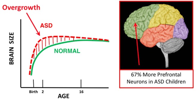
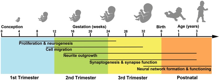
Strong evidence that ASD begins in the womb comes from observations that young ASD male children have 67% more prefrontal neurons than controls (Fig. 1). Neuron overabundance is not uniform in prefrontal cortex: Neuron excess was 79% in dorsolateral and 29% in medial prefrontal cortex. Moreover, the degree of neuron excess across ASD cases was heterogeneous, ranging from 12 to 106%. In four ASD cases, neuron excess was >83% of control average. One child had an excess of 106% and mutation of PTEN, a high-confidence ASD gene. In another study, cortex in young ASD cases had 36% more neurons than controls (Table S1). Young ASD males have ~53% more von Economo neurons in frontoinsular cortex than controls. Cell cycle, differentiation, and DNA damage response genes and pathways are dysregulated in prefrontal cortex at young ages in ASD, and immune gene expression is upregulated. Because proliferation of cortical neurons is exponential between 10 and 20 weeks of gestation and does not occur postnatally, pathological neuron excess cannot be caused by post-natal events and instead indicates neural disorder in ASD has a prenatal origin, likely by the first and/or second trimester.
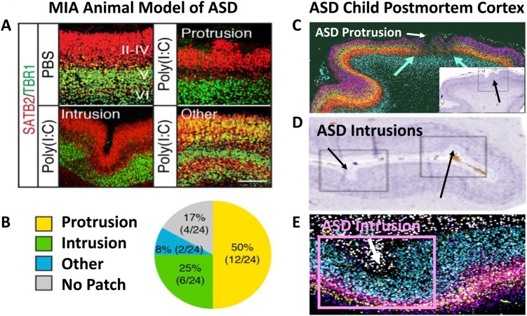
In ASD, neural migration and cortical laminar organization, stages that occur in second and third trimesters, are also abnormal (Fig. 2). Patches of focal laminar disorganization and nearby clusters of mis-migrated neurons were found in prefrontal and temporal cortices in 10 of 11 young ASD cases. In these cases, there was heterogeneity in cortical cell types and layers that were most disrupted. While all layers were affected, patches tended to be pronounced in layers 2, 3 and 4. Patches of cortical dysplasia could be due to somatic mutations. Somatic mutations (e.g., in the PI3K/AKT/mTOR pathway) also occur in hemi-megaencephaly and intractable epilepsy and may be an important prenatal source of phenotypic heterogeneity and risk in ASD and other neurodevelopment disorders.
Neuropathology examination of 4-year-old to 60-year-old ASD brains shows evidence of multiregional dysregulation of neurogenesis and neuronal migration in several cases, including subcortical, periventricular, hippocampal and cerebellar heterotopias reflecting abnormal neuronal migration and multifocal cerebral dysplasias. Complete removal of the subplate, which normally occurs in the third trimester, may also be disrupted in ASD. Reduced growth of neuronal cell size and dendritic arbors in ASD postmortem cases have long been reported and are now recognized as an important neuropathological attribute of ASD cortex; neuron size reduction varies from −4% in occipital to −18% in prefrontal cortex.
ASD patient-derived iPS cells
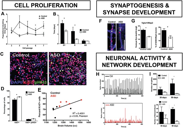
Multiple subsequent fetal age processes were also abnormal in every ASD case in Marchetto et al. ASD patient-derived neurons additionally displayed fewer excitatory synapses, and downregulation of multiple neurotransmitter and synapse markers including GABA receptor markers (Fig. 3f, g). With increasing age from 30 to 50 days of development, ASD-derived neural networks showed less spontaneous excitatory activity and poorly synchronized activity. By 50 days, synchronized neural bursts of activity were 6 times lower in ASD than controls (Fig. 3h, i). The ASD subject with the greatest cell proliferation and brain size has a PTEN mutation and is not intellectually disabled but has severe social communication symptoms. Mariani et al. also reported multiple disrupted fetal stages in their ASD iPSC study, and interestingly these included excessive synaptogenesis. Thus, ASD patient-derived iPSC studies find evidence of multiple fetal stage defects with commonality in excess proliferation and cell cycle acceleration defects, but heterogeneity in synaptogenesis effects. This points to fetal stage subtypes in ASD.
These ASD Living Biology experiments suggest that neural developmental disorder in ASD spans prenatal and early post-natal life, beginning with dysregulation of cell cycle G1/S and excess cell proliferation and ending with disrupted synaptogenesis and dysfunctional nascent neural circuit activity. Heterogeneity already exists at these early stages. Mechanisms that dysregulate proliferation may dysregulate neural differentiation, cell fate determination and maturation. The conclusion that dysregulation of prenatal cell cycle G1/S underlies excess proliferation and early brain overgrowth in these toddlers with ASD is strongly supported by these ASD patient-derived iPS cell data.
Transcriptomic evidence in living ASD toddlers points to prenatal periods
Transcriptomics of leukocytes from ASD toddlers reveal further insights into ASD Living Biology. In analyses of gene expression and MRI measures of brain size at young ages, ASD toddlers with the most abnormal expression of cell cycle hub genes had the most brain overgrowth, while those with milder cell cycle disruption had smaller than normal brain size. Functional genomic analyses identified 23 candidate genes for brain maldevelopment linked to 32 upstream high-confidence ASD genes. Dysregulation of cell cycle G1/S was identified as contributing, suggesting shortened G1/S in bigger ASD brains, consistent with Marchetto et al. CHD8 is one of the most commonly found high-confidence ASD mutations, and the analyses of a CHD8 subnetwork and altered CHD8-regulated transcript levels further confirmed the central role of genes regulating neurogenesis and cell adhesion processes in ASD brain maldevelopment. Thus, in living ASD infants and toddlers, patient-derived iPS cell experiments and leukocyte transcriptomics point to dysregulation of cell cycle G1/S.
High-confidence ASD genes and prenatal development
In a small percentage of children with ASD, rare de novo gene mutations are risk factors. Genetic information accumulated via these and many other studies enable the search for likely ASD genes and gene networks. Unraveling how these mutations affect brain development is crucial to advance understanding of how genetic, non-genetic and gene/environment interactions cause ASD and influence neural development, clinical progression, and treatment. However, new evidence shows many proposed gene associations may be “noise”. Of the hundreds of potential associations, 38 genes are classified as recurrent and potentially penetrant in ASD. Of these 38 genes, 31 also overlap with the 65 SFARI Level 1(S) and 2(S) ASD genes; taken together, these 72 genes are high-confidence ASD (hcASD) genes.
These 72 hcASD genes, many of which regulate gene expression, are the most penetrant genes implicated in ASD thus far. It is crucial to understand how they contribute to brain growth and function because they may point to processes central to ASD development. Previous analyses of ASD risk genes were performed when knowledge of hcASD genes was more limited, and some of those analyses gave similar weight to both low and high-confidence genes. Thus, the developmental roles of these 72 hcASD genes may have been obscured by inclusion of low confidence genes; so further study of these hcASD genes is needed.
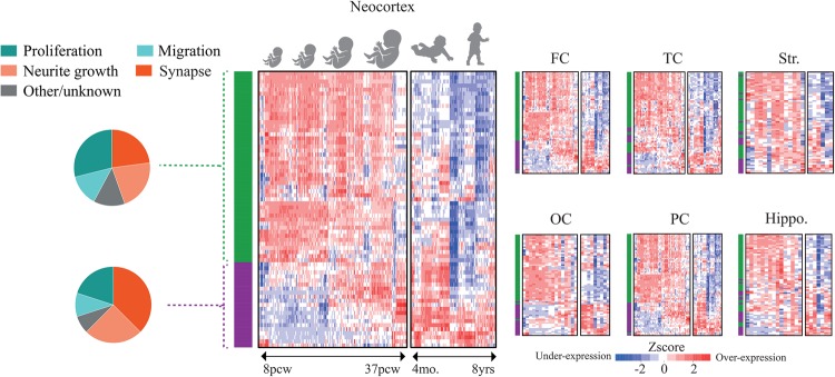
Here we use these 72 hcASD genes to address three central questions: When during development do hcASD genes play developmental roles? Where in the brain do they do so? What functional roles may they play? First, we addressed the “when and where” roles of these hcASD genes by leveraging the most recent BrainSpan RNA-Seq dataset (http://www.brainspan.org/). Of the 72 genes, 3 are not detected (RPKM > 0.5) in >10% of BrainSpan samples between prenatal and up to 8 years old post-natal. Therefore, here we focus on the 69 hcASD genes that do have reliable expression levels.
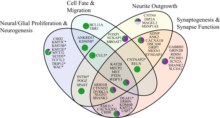
While our analysis highlights the roles of several hcASD genes, it is expected that further work will continue to propose and validate new genes. For example, a recent machine learning approach leveraged previously implicated genes to generate predictions of plausible new autism risk genes. They further demonstrated that these genes converge on fetal and early infancy stages, and were involved in processes including cell cycle, embryogenesis, morphogenesis, axonogenesis and synaptogenesis. Furthermore, these genes also affect multiple brain structures, and are implicated in G1/S, RAS/ERK-PI3K/AKT signaling, and chromatin remodeling.
In summary, our analyses of hcASD genes (Fig. 4) and emerging work on newly predicted ASD risk genes highlight two major human neural development epochs. First, these genes are highly active during early fetal stages of cell proliferation, differentiation, migration and early organization. Second, they impact late fetal generation of synapses and functional assemblies that continue on through birth and impact the early experience and learning years. Thus, hcASD gene evidence dovetails neatly with ASD patient-derived iPS cell and leukocyte transcriptomic evidence in ASD toddlers, and ASD postmortem evidence reviewed above.
Pleiotropic common pathways implicated in ASD
| Gene name | Pathway involved | Examples of effects on pathways |
|---|---|---|
| FMR1 b,c,d | PI3K/AKT | Fmr1 knock-out mice have elevated activation of PI3K/AKT signaling pathway, affecting synaptic plasticity with behavioral and cognitive defects. Downregulation of PI3K in prefrontal cortex of Fmr1 null mice ameliorates aberrations in protein synthesis, dendritic spine density and cortical network activity. |
| FOXG1 b | PI3K/AKT | The PI3K/AKT signaling pathway affects cell fate determination and neural migration by regulating FOXG1. |
| NLGN3 a,c,d | PI3K/AKT | NLGN3 secretion by active neurons enhances proliferation of glial cells by activation of PI3K/AKT pathway. Also affects synaptic development. |
| PTEN a,b,c,d | PI3K/AKT | Pten+/- mice show high activity in PI3K/AKT and β-catenin signaling resulting in enhanced proliferation of radial glial cells and cortical overgrowth; neuronal hypertrophy, hypertrophic and ectopic dendrites and axonal tracts; increased synapses; and hyperconnectivity of prefrontal cortex with amygdala. Pten also involves in neural migration. |
| RELN b,c,d | PI3K/AKT | PI3K/AKT signaling pathway affects cell fate determination and neural migration by regulating RELN. While RELN promotes neuronal maturation, synaptic formation and plasticity, it appears to be more important for dendrite and spine development via PI3K/AKT/mTOR pathway. |
| SHANK3 a,c,d | PI3K/AKT | Mice with Shank3 mutations show deficits in synaptic function, hippocampal LTP and motor performance that can be improved with IGF1 treatment. Also affects neurogenesis. |
| MECP2 a,b,c,d | PI3K/AKT and RAS/ERK | iPS-derived RG cells from Rett syndrome patients with MECP2 mutation exhibit excess proliferation through activation of PI3K/AKT and downregulation of RAS/ERK pathways. Neurons with MECP2 knock-down fail to attain a unipolar/bipolar shape, display abnormal migration and reduced cortical thickness. Neuron models of MECP2 mutations lead to abnormalities in soma size, dendritic arborizations, spine density, and neuronal firing that may be partially rescued by inhibiting Pten gene or IGF1 treatment. |
| ARID1B a,c,d | PI3K/AKT, WNTand β-catenin | ARID1B suppression delays cell cycle re-entry. Mice with Arid1b knock-down show suppressed IGF1, a mediator of PI3K/AKT and β-catenin pathways, resulting in decreased dendritic arborization and accumulation of aberrant dendritic spines and altered synaptic transmission. |
| 16p11.2a,d | RAS/ERK | 16p11.2 is one of most recurrent CNVs in ASD and encompasses multiple genes including ERK1. Its deletion dysregulates RAS/ERK with effect on proliferation and neurogenesis of murine neural progenitor cells. 16p11.2 mutation mouse models show reduced RAS/ERK activity, reduced protein synthesis and cognitive impairments. |
| ERBIN a,d | RAS/ERK | ERBIN has an inhibitory effect on RAS/ERK signaling pathway. Upregulation or downregulation of Erbin leads to enhanced or decreased differentiation of PC12 neurons, respectively. |
| KAT2B a,b,c,d | RAS/ERK | Null mutations lead to dysregulation of RAS/ERK and disruption of the pyramidal cell layer organization. |
| NF1 d | RAS/ERK | Mice with Nf1 null mutations show dysregulated RAS/ERK pathway and deficits in spine morphology, glutamate and GABA release, hippocampal LTP and learning abilities. |
| SYNGAP1 c,d | RAS/ERK | SYNGAP1 acts as RAS inhibitor, and heterozygote mutations in mice show premature neurons, elevated excitatory synaptic transmission, reduced axonal branching and synaptic boutons in inhibitory neurons, and deficits in behavior and cognition. Rescuing Syngap1 in adulthood did not benefit the mice, underscoring its role in prenatal and early post-natal brain development. |
| CHD8 a,d | WNT and β-catenin | CHD8 knock-down disrupts G1/S phase, dysregulates proliferation in neural progenitor and stem cells and causes brain overgrowth. Chd8 mutations in mice result in abnormalities in striatal circuitry and synaptic physiology |
| CTNND2 a,c,d | WNT and β-catenin | Affects proliferation of glioma cells and involved in glioblastoma. Regulates spine morphology; mutations decrease spines and excitatory synapses in hippocampal neurons in rodents. Ctnnd2 mutation in mouse models have impaired spatial learning and fear conditioning. |
Dysregulation of proliferation and neurogenesis
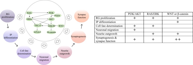
Multiple ASD risk factors, including PTEN, MECP2, CHD8, ARID1B, ERBIN, and the 16p11.2 locus, link ASD to the disruption of neuron production through the PI3K/AKT, RAS/ERK, WNT and β-catenin pathways (Table 1). In precursor and radial glial cells (RGCs), upregulation of PI3K/AKT, WNT and β-catenin or downregulation of RAS/ERK leads to elevated proliferation. The role of these pathways reverses in intermediate progenitor cells (IPCs). Activity of β-catenin (downstream of PI3K/AKT and WNT, Fig. 6) enhances premature differentiation of neurons, while activity of RAS/ERK reduces neurogenesis of IPCs and increases astrogenesis. During human brain development, precursors differentiate into neurons first and glial cells later. Therefore, dysregulation of proliferating precursor cells will perturb the number of generated neurons as well as the balance between neurogenesis and gliogenesis in the developing brain. One of the primary mechanisms that regulate neuroprogenitor divisions is the rate of cell cycle progression, particularly G1/S transition time. Studies on CHD8 and iPS-derived ASD neurons indicate the dysregulation of proliferation and macrocephaly in ASD could be linked to the disruption of these signaling pathways and shortened G1/S transition. Importantly, the induced proliferation by the signaling pathways can result in either a thicker or thinner cortex in mice models depending on the strength of the dysregulation. While downregulation of RAS/ERK can lead to a thicker cortex from increased proliferation and an increased neuronal count, loss of Erk2 can induce such strong G1/S dysregulation that mice cells miss the time window of neurogenesis (E14.5–E16.5), resulting in fewer neurons, precocious astrogenesis, and potentially microcephaly. The other key factor that controls neuron numbers is the balance between cell cycle re-entry (proliferative divisions) and exit (neurogenesis). During brain development, RGCs can divide symmetrically or asymmetrically. While symmetric divisions result in two daughter RGCs, asymmetric divisions produce a RGC and one another cell. The newly generated cell can differentiate into a neuron, or become an IPC. IPCs can undergo 1–3 proliferation cycles before differentiation to neurons. Cortical surface area and thickness are controlled by the mechanisms involved in RGC and IPC proliferation and differentiation decisions. Interestingly, G1/S transition rates might be associated with these cell decisions as symmetric divisions can have shorter G1/S transition times than asymmetric divisions. Evidence suggests that PI3K/AKT, RAS/ERK, WNT and β-catenin pathways are major players of these cell decisions as well. RGCs in a mouse model of Gsk3 knockout, a major convergence point of the signaling pathways (Fig. 6), show more highly active β-catenin and Notch pathways. As a result, RGCs were locked in proliferative state with attenuated neurogenesis. This resulted in expanded RGC pools, fewer IPCs, reduced cortical thickness and increased ventricular apical surface length. Hence, it is tempting to speculate that perturbation of these four pathways in ASD could involve dysregulation of balance between proliferation and neurogenesis as well as neurogenesis and gliogenesis.
Disruption of cell fate and migration
Studies on MECP2, KAT2B, REELIN and FOXG1 hcASD genes suggest that they are associated with changes to the cell fate determination and neural migration processes in ASD through PI3K/AKT, RAS/ERK, WNT and β-catenin pathways (Table 1). Upregulation of WNT and β-catenin signaling, for example, prevents the multipolar to bipolar transition of pyramidal precursors. Thus, neurons fail to migrate to the cortical plate and accumulate within the intermediate zone. Somatic mutations can also lead to dysregulation of these signaling pathways during brain development. Somatic activating mutations in the PI3K/AKT pathway are associated with hemimegalencephaly and focal cortical dysplasia syndromes that are marked by cortical dyslamination, dysmorphic neurons and loss of radial neuronal orientation. Thus, in addition to modulating neuron development, the perturbed pathways in ASD also alter cell fate determination and neural migration.
Dysregulation of neurite outgrowth and neuronal function
The hcASD genes SHANK3, FMR1, CTNND2, CHD8, SYNGAP1, MECP2, PTEN, ARID1B and NF1 are associated with dysregulation of neurite outgrowth and neuronal function through the PI3K/AKT, RAS/ERK, WNT and β-catenin signaling pathways (Table 1). These pathways control soma size, dendritic arborization, axon generation, spine development and synapse function. Activity of receptor tyrosine kinases, metabotropic glutamic receptors and NMDA receptors lead to the activation of PI3K/AKT and RAS/ERK signaling pathways, and thereby influence synaptic plasticity by mediating expression and trafficking of AMPA receptors in excitatory synapses and controlling long-term depression (LTD) and long-term potentiation (LTP) responses. PI3K/AKT and RAS/ERK signaling pathways also control processes associated with neural excitotoxicity and cell survival. Interestingly, it seems that hcASD genes do not impact these pathways in the same way. For example, while mutations in Fmr1 and Pten lead to the over-activity of PI3K/AKT pathway with increased synaptogenesis and hyperconnectivity of the neurons, mutations in Mecp2 and Shank3 are reported to suppress the pathway, leading to delayed and sparse spine development and reduced synaptic amplitude. Interestingly, in cases of Mecp2 and Shank3, neurons could be partially rescued by activating PI3K/AKT pathway through Pten inhibition or IGF1 treatment, while downregulation of the pathway in Fmr1 null mice ameliorated aberrations in protein synthesis, dendritic spine density and cortical network connectivity.
Finally, by prenatal injection of a small molecule that stabilizes axin, a component of the WNT and β-catenin signaling pathway, cell cycle proliferation is prolonged and causes an excess of layer 2 and 3 neurons and early brain overgrowth, abnormal synaptogenesis and increased excitatory neural activity. This was accompanied by ASD-like abnormal social, vocalization, and ritualistic behaviors. These results strengthen the causal link between cell cycle disruption and ASD and also suggest that non-genetic etiologies may underlie deviant neurodevelopmental trajectories.
Thus, RAS/ERK, PI3K/AKT, WNT and β-catenin are highly integrated and pleiotropic, exhibiting different and successive roles throughout prenatal and early post-natal life (Fig. 6). Across ASD individuals, variation in how and when these pleiotropic pathways are dysregulated will cause early heterogeneity of pathophysiology and potentially later neural and clinical heterogeneous outcomes.
The prenatal maternal immune activation model of ASD
Non-genetic etiologies may account for 30–41% of the risk for ASD, and such factors may act independently or in combination with genetic factors. A well-established non-genetic model system for studying ASD pathophysiology is the prenatal maternal immune activation (MIA) model. The link between MIA and enhanced risk for ASD is bolstered by large-scale population-based studies showing small but significantly increased risk due to maternal prenatal infections. MIA models have ASD-like social, vocalization, ritualistic, exploration and other behavioral deficits. Current MIA ASD models expose developing rodent pups to bacterial or viral mimetics to elicit strong immune responses. This is timed to roughly mimic infection between 8 to 12 weeks during the 1st trimester in humans, when neural progenitor cells are proliferating to expand cortical surface area and promote cortical layering.
| Human development | Abnormalities induced by MIA and associated with ASD |
|---|---|
| 1st–2nd Trimester | Upregulated cell cycle and downregulated migration and neurite outgrowth gene expression |
| Cortical layering: over-production of neurons, increased cortical thickness, focal cortical dysplasia | |
| Cerebellar vermis dysplasia | |
| 2nd–3rd Trimester | Microglia: enhanced priming, activation |
| Dendritic morphology abnormalities | |
| GABAergic signaling, excitatory/inhibitory imbalance, number of interneurons | |
| 3rd Trimester—post-natal | White matter neuron density |
| Dendritic spines number and turnover rates | |
| Synaptic pruning and proteins | |
| Post-natal | Early brain overgrowth |
| Myelin functionality and stability | |
| Dopamine system | |
| Serotonin levels | |
| ASD-like abnormal social, vocalization, and ritualistic behaviors | |
| Gender-dependent effects | |
| Transgenerational effects |
Given the diverse ASD-associated phenotypes that arise with a prenatal immune challenge, MIA-induced effects on neurodevelopment provide insights into the molecular mechanisms behind ASD and related neurodevelopmental disorders. Within the ASD neuropathological and molecular heterogeneous landscape, MIA disrupts fetal development by involving transcriptional programs that are abnormal in adult ASD brain tissue and genes with high-confidence ASD mutations or their downstream targets. This evidence suggests shared disrupted pathways between genetic and non-genetic etiologies and the possibility for a much more relevant interplay between a genetic background at-risk for ASD and environmental triggers. Work by Le Belle et al. identified that brain enlargement due to Pten haploinsufficiency in mouse increased from 8 to 44% when combined with MIA, thus demonstrating the interactive effects of MIA and ASD gene mutations in the disruption of cortical development. Such augmented disruption occurs via the activation of the cellular redox signaling in response to infection. Reactive oxygen species (ROS) can increase stem cell self-renewal and neurogenesis through the reversible inactivation of PTEN and hyper-activation of the NADPH oxidase (NOX)-PI3K pathway, which ultimately, during early post-natal development, will result in enlarged brains and autistic behaviors.
MIA also causes long-lasting deleterious effects on the post-natal brain similar to those expected from germline mutations in patients with neurodevelopmental disorders. Such effects likely involve the epigenetic dysregulation of transcriptional programs that are sensitive to environmental changes or insults. Inflammatory mediators (e.g., cytokines) induce such alterations during neurodevelopment by histone modifications, methylation changes at the promoter of ASD-critical genes (e.g., MECP2) or genes involved in synaptic functions and global methylation changes. Importantly, increased levels of IL-6 alone recapitulate aspects of ASD pathophysiology. At the molecular level, IL-6 increases the activity of DNA methyltransferases 1 (DNMT1) with activating effects on the JAK/STAT3, NF-κB and PI3K/AKT/mTOR signaling pathways affecting the regulation of autophagy-related functions, that are required to maintain cellular homeostasis under stress, such as a prenatal insult. This provides a strong link between pro-inflammatory immune factors, body growth through activation of the mTOR pathway and uncontrolled proliferation typical of cancer-related disease. Genetic or epigenetic dysregulation of such pathways is associated with several neurodevelopmental disorders, in particular psychiatric, characterized by abnormal brain size (enlarged and reduced). Consistently, we found that MIA alters key components of the TSC/mTOR signaling pathway with ultimate activation of the translation initiation factor EIF4E which is predicted to alter the regulation of neural progenitor cell divisions during midgestation. Such regulation may also be altered by activation of microglia cells under stress conditions including MIA and/or in response to increase levels of IL-6. Impairment in the regulation of microglia functions is consistently seen in ASD studies and is likely involved in abnormal regulation of neuron numbers both at pre- and post-natal stages.
Accumulating evidence consolidates the idea that environmental models of ASD-associated phenotypes may provide valuable insights to help develop potential treatment, prevention, or intervention strategies. For example, MIA alters expression in pathways shared with those in fragile X syndrome for which advances in drug development are in progress. With a better understanding of mechanisms behind environmental etiologies of ASD, such as MIA, we could potentially identify therapeutic targets amenable to prevention and/or treatment later in life. In this scenario, drugs that successfully target those pathways in fragile X syndrome could potentially be re-purposed. Other studies have used mesenchymal stem cells or pharmacological treatment to block or antagonize the effect of MIA on purinergic or inflammatory pathways.
Future directions: ASD Living Biology
Evidence shows that ASD begins during prenatal life, which is the least studied and understood of all developmental periods of ASD. This glaring gap poses a major barrier to progress in ASD research and treatment and exists because nearly all current approaches—postmortem, molecular, genomic, genetic, computational and animal models—cannot get answers to foundational questions such as: What are the underlying molecular and cellular processes during fetal and early post-natal stages that cause ASD in each child, and how does this vary across children? How do different prenatal trajectories explain and predict heterogeneity in post-natal brain growth and functioning, behavior and clinical profiles in ASD toddlers? Are there subtypes that display normal fetal-stage development but begin abnormal development at post-natal stages?
Nor can current approaches effectively examine more targeted questions such as: How do different hcASD genes affect fetal processes, and how are those effects related to post-natal cognitive, language and social development? Is there a difference in the brain development trajectory between idiopathic ASD cases and ASD with mutations in hcASD risk genes? Since macrocephaly is more strongly related to ASD than to intellectual disability (ID) and many hcASD genes such as CHD8 and WDFY3 cause dysregulation of proliferation and brain overgrowth, what other fetal mechanisms are altered by excess proliferation that lead to an ASD outcome? What are the fetal-stage processes that alter imbalance between excitation and inhibition that is proposed to explain learning, memory, sensorimotor, and cognitive deficits in ASD ?
Post-natal phenotype data alone cannot reveal fetal-stage biology, and fetal-stage cellular models alone cannot explain post-natal development and clinical heterogeneity. There is a need to bridge the gap between fetal and post-natal biology for living ASD toddlers, and this requires a paradigm shift. This paradigm we describe here, ASD Living Biology, approaches the fundamental questions above, by acquiring fetal and early post-natal measures using iPS cells derived from skin or blood at any age, and integrating them using a within-subjects design. Discovery of the subtypes of prenatal pathogenic processes and their interplay with post-natal experiences should be a major direction for ASD research.
The ASD Living Biology approach will enable the construction of quantitative multiscale models that could address the perturbations in fetal-to-post-natal development. These explanatory and predictive models of ASD could address the disorder at the individual, subtype and group level, when compared to models from typically developing children. ASD Living Biology models would link in vitro and in vivo data. In vitro data would include molecular, cellular and physiological measures of a child’s iPS cells, including measures of fetal-stage proliferation/differentiation, cell fate and growth, synaptogenesis, synaptic function and synchronized neural network activity. This iPSC based approach could then be connected with in vivo measurements from the same child, including their neural structural and functional, genetic and genomic, behavioral, psychometric, diagnostic and clinical outcome data. By thus integrating such within-child fetal and post-natal measures, and integrating these data, it will be possible to better understand prenatal molecular mechanisms leading to the development of ASD.
Interactions of post-natal biology and experiences with preceding prenatal biology
Future research could also determine whether some ASD prenatal trajectories are more deterministic of outcome phenotype, while others might be easily modified by experience, learning and interventions. The pleiotropic effect of many hcASD genes raises the testable hypothesis of a layered prenatal to post-natal path toward ASD in which the disorder may be dynamically impacted by post-natal changing functional roles of some ASD genes. It is also possible that heterogeneity in ASD outcomes may derive from the interplay of diverse prenatal dysregulations with post-natal exposures. Post-natal changes are thought to be reflected in changing behavioral and neurodevelopmental features and some underlying mechanisms have been proposed. Perhaps different prenatal pathogenic subtypes act as different general starting points from which diverse post-natal experiences lead to further variation in atypical development, some to higher and others to lower risk for ASD. For each child, prenatal starting points must be “worked around” to find optimal adaptations for that individual. As a result, more or less severe atypical behavior and cognitive outcomes may occur. ASD Living Biology studies could be designed to look at the interaction between atypical prenatal biology and post-natal experience-based adaptation and how this could lead to a spiral in early development that canalizes over time toward multiple, heterogeneous end points (e.g., multifinality). Because neural circuits are sculpted by experience, future designs could examine whether atypical reduced social visual engagements from very early in life in ASD, are the product of some atypical prenatal to post-natal neural pathophysiology that causes an individual to engage in behavioral and cognitive adaptations that result in decreased sampling of the important social information necessary for typical social brain development. The more atypical such post-natal experience becomes, the more an individual’s development could become canalized for atypical outcomes.
Conclusions
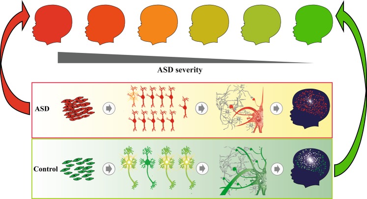
ASD is a multi-etiology, multi-stage, progressive disorder that spans most of fetal life. In our theory, a single perturbation can trigger a cascade of pathogenic changes, such as an MIA event, a single fetal injection of XAV939 or PTEN mutation in a child. Multiple perturbations may substantially amplify both the severity of disorder and outcome heterogeneity, such as, for example, a combination of PTEN mutation and MIA. Moreover, many known and proposed ASD etiologies (e.g., MECP2, PTEN, MIA, XAV939) may perturb the pleiotropic RAS/ERK-PI3K/AKT pathway (Fig. 6), which in turn modulates each of the ASD-critical fetal and early post-natal stages discussed here. Dysregulated proliferation/differentiation can be a potent perturbation because it can lead to abnormal changes in subsequent stages of cell fate, migration, organization, maturation, and synapse and neural development. Cell and animal models of high-confidence ASD genes, such as CHD8, PTEN, WDFY3, definitively show disruption of cell proliferation (Fig. 5; Table S2); Fang et al. show robust synaptic, neural and behavioral downstream effects of excess neuron proliferation, and ASD patient-derived iPSC models show that excess proliferation is a robust early fetal stage defect in every case examined.
Culprit prenatal perturbations do not necessarily produce identical neural and clinical outcomes even when the perturbations are themselves identical in siblings. The Choi and colleagues MIA ASD mouse model study showed that the same fetal MIA perturbation of littermates causes patches of focal cortical dysplasia that have marked heterogeneity, analogous to heterogeneity found in cortex in unrelated individual ASD cases postmortem (Fig. 7). The Wdfy3 mutation mouse model of ASD likewise displays heterogeneity of migration and focal patch defects across individual animals, as well as disrupted proliferation/differentiation and cortical overgrowth. In Courchesne et al., two ASD individuals had >100% the normal mean prefrontal neuron number, but one had small neurons and a modestly enlarged brain weight while the other had typical neuron size and one of the largest brain weights ever recorded for ASD. In the Marchetto et al. study, even though neural progenitors from every ASD toddler doubled cell numbers faster than every control, the proliferation rates among ASD toddlers varied and that variation correlated with variation in MRI volume.
Because ASD begins in prenatal and early post-natal life, there is an ethical demand for early detection, intervention and services. When it is not detected in an infant or toddler, it is because it was missed. Studies demonstrate that ASD risk can be measured as young as 12 to 24 months using parent report screening tools such as the CSBS IT-Checklist and M-CHAT at well-baby check-ups with follow-up confirmation at ASD specialty clinics. This screening procedure is fast, easy, inexpensive and effective, and can be done in any pediatric office or clinic. Studies show that when pediatricians and ASD specialty clinics work together, risk detection and diagnostic evaluation can occur as early as 12 to 20 months, and interventions soon thereafter.
Early interventions and services may improve an ASD child’s developmental outcome and help parents at a crucial time in human brain development. During the first post-natal years, the human brain undergoes a profound period of establishing and refining neural connections, and this is the basis for the emergence of higher-order social, language and cognitive networks and behavior. This important developmental step of the construction of functional and adaptive neural circuits is dependent on adaptive neural responses to input from the environment. If an infant or toddler with ASD is identified and behavioral treatment begun before or while early brain connections are being actively established, then brain function for that toddler stands the best chance of being improved. This is superior to treatment that begins late and after abnormal mature circuitry is already established. It is for this very reason that the early identification and treatment of ASD is essential and ethically demanded.
Lastly, to address the progressive prenatal beginnings and likely prenatal to post-natal experience-dependent interactions in ASD, new treatment designs are needed. We advocate coordinated in vitro and in vivo within-subject ASD Living Biology research designs to provide early-age molecular, cellular, and neuronal functional developmental explanations for individual differences as well as across ASD commonalities. Advances in stem cell model systems enable this: Patient-derived brain organoids allow investigation of fetal processes that are remarkably similar to in vivo development. Within-child in vitro and in vivo designs could reveal prenatal and early post-natal bases of outcome clinical heterogeneity. Through this ASD Living Biology approach, relationships between prenatal development and post-natal experience, learning, brain growth and function, clinical presentation and progression, and treatment responsiveness may be defined at the individual level, thus enabling the development of precision medicine approaches with truly beneficial interventions.
Sources
- Kanner LAutistic disturbances of affective contactNerv Child1943221750 https://pubmed.ncbi.nlm.nih.gov/4880460/
- Constantino JN, Marrus NThe early origins of autismChild Adolesc Psychiatr Clin N Am20172655570 https://doi.org/10.1016/j.chc.2017.02.008
- Dawson G, Meltzoff AN, Osterling J, Rinaldi J, Brown EChildren with autism fail to orient to naturally occurring social stimuliJ Autism Dev Disord19982847985 https://doi.org/10.1023/a:1026043926488
- Lord C, Bishop S, Anderson DDevelopmental trajectories as autism phenotypesAm J Med Genet C Semin Med Genet2015169198208 https://doi.org/10.1002/ajmg.c.31440
- Courchesne E, Karns C, Davis HR, Ziccardi R, Carper R, Tigue ZUnusual brain growth patterns in early life in patients with autistic disorder: An MRI studyNeurology20015724554 https://doi.org/10.1212/wnl.57.2.245
- Courchesne E, Pierce KWhy the frontal cortex in autism might be talking only to itself: local over-connectivity but long-distance disconnectionCurr Opin Neurobiol20051522530 https://doi.org/10.1016/j.conb.2005.03.001
- Courchesne E, Pierce K, Schumann CM, Redcay E, Buckwalter JA, Kennedy DPMapping early brain development in autismNeuron200756399413 https://doi.org/10.1016/j.neuron.2007.10.016
- Carper RA, Moses P, Tigue ZD, Courchesne ECerebral lobes in autism: early hyperplasia and abnormal age effectsNeuroimage200216103851 https://doi.org/10.1006/nimg.2002.1099
- Carper RA, Courchesne ELocalized enlargement of the frontal cortex in early autismBiol Psychiatry20055712633 https://doi.org/10.1016/j.biopsych.2004.11.005
- Sparks BF, Friedman SD, Shaw DW, Aylward EH, Echelard D, Artru AABrain structural abnormalities in young children with autism spectrum disorderNeurology20025918492 https://doi.org/10.1212/wnl.59.2.184
- Hazlett HC, Poe M, Gerig G, Smith RG, Provenzale J, Ross AMagnetic resonance imaging and head circumference study of brain size in autism: birth through age 2 yearsArch Gen Psychiatry200562136676 https://doi.org/10.1001/archpsyc.62.12.1366
- Stanfield AC, McIntosh AM, Spencer MD, Philip R, Gaur S, Lawrie SMTowards a neuroanatomy of autism: a systematic review and meta-analysis of structural magnetic resonance imaging studiesEur Psychiatry20082328999 https://doi.org/10.1016/j.eurpsy.2007.05.006
- Shen MD, Nordahl CW, Young GS, Wootton-Gorges SL, Lee A, Liston SEEarly brain enlargement and elevated extra-axial fluid in infants who develop autism spectrum disorderBrain2013136Pt 9282535 https://doi.org/10.1093/brain/awt166
- Schumann CM, Bloss CS, Barnes CC, Wideman GM, Carper RA, Akshoomoff NLongitudinal magnetic resonance imaging study of cortical development through early childhood in autismJ Neurosci201030441927 https://doi.org/10.1523/JNEUROSCI.5714-09.2010
- Zielinski BA, Prigge MB, Nielsen JA, Froehlich AL, Abildskov TJ, Anderson JSLongitudinal changes in cortical thickness in autism and typical developmentBrain2014137Pt 61799812 https://doi.org/10.1093/brain/awu083
- Lange N, Travers BG, Bigler ED, Prigge MB, Froehlich AL, Nielsen JALongitudinal volumetric brain changes in autism spectrum disorder ages 6-35 yearsAutism Res201588293 https://doi.org/10.1002/aur.1427
- Ohta H, Nordahl CW, Iosif AM, Lee A, Rogers S, Amaral DGIncreased surface area, but not cortical thickness, in a subset of young boys with autism spectrum disorderAutism Res2016923248 https://doi.org/10.1002/aur.1520
- Hazlett HC, Gu H, Munsell BC, Kim SH, Styner M, Wolff JJEarly brain development in infants at high risk for autism spectrum disorderNature201754234851 https://doi.org/10.1038/nature21369
- Solso S, Xu R, Proudfoot J, Hagler DJ, Campbell K, Venkatraman VDiffusion tensor imaging provides evidence of possible axonal overconnectivity in frontal lobes in autism spectrum disorder toddlersBiol Psychiatry20167967684 https://doi.org/10.1016/j.biopsych.2015.06.029
- Courchesne E, Carper R, Akshoomoff NEvidence of brain overgrowth in the first year of life in autismJAMA200329033744 https://doi.org/10.1001/jama.290.3.337
- Dementieva YA, Vance DD, Donnelly SL, Elston LA, Wolpert CM, Ravan SAAccelerated head growth in early development of individuals with autismPediatr Neurol2005321028 https://doi.org/10.1016/j.pediatrneurol.2004.08.005
- Dawson G, Munson J, Webb SJ, Nalty T, Abbott R, Toth KRate of head growth decelerates and symptoms worsen in the second year of life in autismBiol Psychiatry20076145864 https://doi.org/10.1016/j.biopsych.2006.07.016
- Redcay E, Courchesne EWhen is the brain enlarged in autism? A meta-analysis of all brain size reportsBiol Psychiatry20055819 https://doi.org/10.1016/j.biopsych.2005.03.026
- Courchesne E, Mouton PR, Calhoun ME, Semendeferi K, Ahrens-Barbeau C, Hallet MJNeuron number and size in prefrontal cortex of children with autismJAMA2011306200110 https://doi.org/10.1001/jama.2011.1638
- Bauman ML, Kemper TLNeuroanatomic observations of the brain in autism: a review and future directionsInt J Dev Neurosci2005231837 https://doi.org/10.1016/j.ijdevneu.2004.09.006
- Levy J, Coussement A, Dupont C, Guimiot F, Baumann C, Viot GMolecular and clinical delineation of 2p15p16.1 microdeletion syndromeAm J Med Genet A201717320817 https://doi.org/10.1002/ajmg.a.38302
- Bernier R, Steinman KJ, Reilly B, Wallace AS, Sherr EH, Pojman NClinical phenotype of the recurrent 1q21.1 copy-number variantGenet Med2016183419 https://doi.org/10.1038/gim.2015.78
- Jang DH, Chae H, Kim MAutistic and Rett-like features associated with 2q33.3-q34 interstitial deletionAm J Med Genet A2015167A22138 https://doi.org/10.1002/ajmg.a.37119
- Kumar RA, Sudi J, Babatz TD, Brune CW, Oswald D, Yen MA de novo 1p34.2 microdeletion identifies the synaptic vesicle gene RIMS3 as a novel candidate for autismJ Med Genet2010478190 https://doi.org/10.1136/jmg.2008.065821
- Sacco R, Gabriele S, Persico AMHead circumference and brain size in autism spectrum disorder: a systematic review and meta-analysisPsychiatry Res201523423951 https://doi.org/10.1016/j.pscychresns.2015.08.016
- Donovan AP, Basson MAThe neuroanatomy of autism—a developmental perspectiveJ Anat2017230415 https://doi.org/10.1111/joa.12542
- Packer ANeocortical neurogenesis and the etiology of autism spectrum disorderNeurosci Biobehav Rev20166418595 https://doi.org/10.1016/j.neubiorev.2016.03.002
- Kaushik G, Zarbalis KSPrenatal neurogenesis in autism spectrum disordersFront Chem2016412 https://doi.org/10.3389/fchem.2016.00012
- Stoner R, Chow ML, Boyle MP, Sunkin SM, Mouton PR, Roy SPatches of disorganization in the neocortex of children with autismN Engl J Med2014370120919 https://doi.org/10.1056/NEJMoa1307491
- Gotz M, Huttner WBThe cell biology of neurogenesisNat Rev Mol Cell Biol2005677788 https://doi.org/10.1038/nrm1739
- Bayatti N, Moss JA, Sun L, Ambrose P, Ward JF, Lindsay SA molecular neuroanatomical study of the developing human neocortex from 8 to 17 postconceptional weeks revealing the early differentiation of the subplate and subventricular zoneCereb Cortex200818153648 https://doi.org/10.1093/cercor/bhm184
- Hansen DV, Lui JH, Parker PR, Kriegstein ARNeurogenic radial glia in the outer subventricular zone of human neocortexNature201046455461 https://doi.org/10.1038/nature08845
- Fish JL, Dehay C, Kennedy H, Huttner WBMaking bigger brains-the evolution of neural-progenitor-cell divisionJ Cell Sci2008121Pt 17278393 https://doi.org/10.1242/jcs.023465
- Kriegstein A, Noctor S, Martinez-Cerdeno VPatterns of neural stem and progenitor cell division may underlie evolutionary cortical expansionNat Rev Neurosci2006788390 https://doi.org/10.1038/nrn2008
- van Kooten IA, Palmen SJ, von Cappeln P, Steinbusch HW, Korr H, Heinsen HNeurons in the fusiform gyrus are fewer and smaller in autismBrain2008131Pt 498799 https://doi.org/10.1093/brain/awn033
- Santos M, Uppal N, Butti C, Wicinski B, Schmeidler J, Giannakopoulos PVon Economo neurons in autism: a stereologic study of the frontoinsular cortex in childrenBrain Res2011138020617 https://doi.org/10.1016/j.brainres.2010.08.067
- Chow ML, Pramparo T, Winn ME, Barnes CC, Li HR, Weiss LAge-dependent brain gene expression and copy number anomalies in autism suggest distinct pathological processes at young versus mature agesPLoS Genet20128e1002592 https://doi.org/10.1371/journal.pgen.1002592
- Rabinowicz T, de Courten-Myers GM, Petetot JM, Xi G, de los Reyes EHuman cortex development: estimates of neuronal numbers indicate major loss late during gestationJ Neuropathol Exp Neurol1996553208 https://pubmed.ncbi.nlm.nih.gov/8786390/
- Gohlke JM, Griffith WC, Faustman EMComputational models of neocortical neuronogenesis and programmed cell death in the developing mouse, monkey, and humanCereb Cortex200717243342 https://doi.org/10.1093/cercor/bhl151
- Wegiel J, Kuchna I, Nowicki K, Imaki H, Marchi E, Ma SYThe neuropathology of autism: defects of neurogenesis and neuronal migration, and dysplastic changesActa Neuropathol201011975570 https://doi.org/10.1007/s00401-010-0655-4
- Varghese M, Keshav N, Jacot-Descombes S, Warda T, Wicinski B, Dickstein DLAutism spectrum disorder: neuropathology and animal modelsActa Neuropathol201713453766 https://doi.org/10.1007/s00401-017-1736-4
- Hutsler JJ, Casanova MFReview: Cortical construction in autism spectrum disorder: columns, connectivity and the subplateNeuropathol Appl Neurobiol20164211534 https://doi.org/10.1111/nan.12227
- Bailey A, Luthert P, Dean A, Harding B, Janota I, Montgomery MA clinicopathological study of autismBrain1998121Pt 5889905 https://doi.org/10.1093/brain/121.5.889
- D’Gama AM, Pochareddy S, Li M, Jamuar SS, Reiff RE, Lam ANTargeted DNA sequencing from autism spectrum disorder brains implicates multiple genetic mechanismsNeuron2015889107 https://doi.org/10.1016/j.neuron.2015.11.009
- McConnell MJ, Moran JV, Abyzov A, Akbarian S, Bae T, Cortes-Ciriano I et al. Intersection of diverse neuronal genomes and neuropsychiatric disease: The Brain Somatic Mosaicism Network. Science. 2017;356. https://doi.org/10.1126/science.aal1641
- Avino TA, Hutsler JJAbnormal cell patterning at the cortical gray-white matter boundary in autism spectrum disordersBrain Res2010136013846 https://doi.org/10.1016/j.brainres.2010.08.091
- Raymond GV, Bauman ML, Kemper TLHippocampus in autism: a Golgi analysisActa Neuropathol1996911179 https://doi.org/10.1007/s004010050401
- Jacot-Descombes S, Uppal N, Wicinski B, Santos M, Schmeidler J, Giannakopoulos PDecreased pyramidal neuron size in Brodmann areas 44 and 45 in patients with autismActa Neuropathol20121246779 https://doi.org/10.1007/s00401-012-0976-6
- Marchetto MC, Belinson H, Tian Y, Freitas BC, Fu C, Vadodaria KCAltered proliferation and networks in neural cells derived from idiopathic autistic individualsMol Psychiatry20162282035 https://doi.org/10.1038/mp.2016.95
- Mariani J, Coppola G, Zhang P, Abyzov A, Provini L, Tomasini LFOXG1-dependent dysregulation of GABA/Glutamate neuron differentiation in autism spectrum disordersCell201516237590 https://doi.org/10.1016/j.cell.2015.06.034
- Pramparo T, Lombardo MV, Campbell K, Barnes CC, Marinero S, Solso SCell cycle networks link gene expression dysregulation, mutation, and brain maldevelopment in autistic toddlersMol Syst Biol201511841 https://doi.org/10.15252/msb.20156108
- Krumm N, O’Roak BJ, Shendure J, Eichler EEA de novo convergence of autism genetics and molecular neuroscienceTrends Neurosci20143795105 https://doi.org/10.1016/j.tins.2013.11.005
- O’Roak BJ, Vives L, Girirajan S, Karakoc E, Krumm N, Coe BPSporadic autism exomes reveal a highly interconnected protein network of de novo mutationsNature201248524650 https://doi.org/10.1038/nature10989
- De Rubeis S, He X, Goldberg AP, Poultney CS, Samocha K, Cicek AESynaptic, transcriptional and chromatin genes disrupted in autismNature201451520915 https://doi.org/10.1038/nature13772
- Sanders SJ, Murtha MT, Gupta AR, Murdoch JD, Raubeson MJ, Willsey AJDe novo mutations revealed by whole-exome sequencing are strongly associated with autismNature201248523741 https://doi.org/10.1038/nature10945
- Iossifov I, Ronemus M, Levy D, Wang Z, Hakker I, Rosenbaum JDe novo gene disruptions in children on the autistic spectrumNeuron20127428599 https://doi.org/10.1016/j.neuron.2012.04.009
- Stessman HA, Xiong B, Coe BP, Wang T, Hoekzema K, Fenckova MTargeted sequencing identifies 91 neurodevelopmental-disorder risk genes with autism and developmental-disability biasesNat Genet20174951526 https://doi.org/10.1038/ng.3792
- Iossifov I, O’Roak BJ, Sanders SJ, Ronemus M, Krumm N, Levy DThe contribution of de novo coding mutations to autism spectrum disorderNature201451521621 https://doi.org/10.1038/nature13908
- Krumm N, Turner TN, Baker C, Vives L, Mohajeri K, Witherspoon KExcess of rare, inherited truncating mutations in autismNat Genet2015475828 https://doi.org/10.1038/ng.3303
- O’Roak BJ, Deriziotis P, Lee C, Vives L, Schwartz JJ, Girirajan SExome sequencing in sporadic autism spectrum disorders identifies severe de novo mutationsNat Genet2011435859 https://doi.org/10.1038/ng.835
- Kosmicki JA, Samocha KE, Howrigan DP, Sanders SJ, Slowikowski K, Lek MRefining the role of de novo protein-truncating variants in neurodevelopmental disorders by using population reference samplesNat Genet20174950410 https://doi.org/10.1038/ng.3789
- Willsey AJ, Sanders SJ, Li M, Dong S, Tebbenkamp AT, Muhle RACoexpression networks implicate human midfetal deep cortical projection neurons in the pathogenesis of autismCell20131559971007 https://doi.org/10.1016/j.cell.2013.10.020
- Parikshak NN, Luo R, Zhang A, Won H, Lowe JK, Chandran VIntegrative functional genomic analyses implicate specific molecular pathways and circuits in autismCell2013155100821 https://doi.org/10.1016/j.cell.2013.10.031
- Chao HT, Chen H, Samaco RC, Xue M, Chahrour M, Yoo JDysfunction in GABA signalling mediates autism-like stereotypies and Rett syndrome phenotypesNature20104682639 https://doi.org/10.1038/nature09582
- Zoghbi HYPostnatal neurodevelopmental disorders: meeting at the synapse?Science200330282630 https://doi.org/10.1126/science.1089071
- Li H, Zhong X, Chau KF, Santistevan NJ, Guo W, Kong GCell cycle-linked MeCP2 phosphorylation modulates adult neurogenesis involving the Notch signalling pathwayNat Commun201455601 https://doi.org/10.1038/ncomms6601
- Neul JL, Zoghbi HYRett syndrome: a prototypical neurodevelopmental disorderNeuroscientist20041011828 https://doi.org/10.1177/1073858403260995
- Mellios N, Feldman D A, Sheridan S D, Ip J P K, Kwok S, Amoah S K, Rosen B, Rodriguez B A, Crawford B, Swaminathan R, Chou S, Li Y, Ziats M, Ernst C, Jaenisch R, Haggarty S J, Sur MMeCP2-regulated miRNAs control early human neurogenesis through differential effects on ERK and AKT signalingMolecular Psychiatry201723410511065 https://doi.org/10.1038/mp.2017.86
- Zhang ZN, Freitas BC, Qian H, Lux J, Acab A, Trujillo CALayered hydrogels accelerate iPSC-derived neuronal maturation and reveal migration defects caused by MeCP2 dysfunctionProc Natl Acad Sci USA2016113318590 https://doi.org/10.1073/pnas.1521255113
- Larimore JL, Chapleau CA, Kudo S, Theibert A, Percy AK, Pozzo-Miller LBdnf overexpression in hippocampal neurons prevents dendritic atrophy caused by Rett-associated MECP2 mutationsNeurobiol Dis200934199211 https://doi.org/10.1016/j.nbd.2008.12.011
- Monteiro P, Feng GSHANK proteins: roles at the synapse and in autism spectrum disorderNat Rev Neurosci20171814757 https://doi.org/10.1038/nrn.2016.183
- Zhao H, Tu Z, Xu H, Yan S, Yan H, Zheng YAltered neurogenesis and disrupted expression of synaptic proteins in prefrontal cortex of SHANK3-deficient non-human primateCell Res20172712937 https://doi.org/10.1038/cr.2017.95
- Krishnan A, Zhang R, Yao V, Theesfeld CL, Wong AK, Tadych AGenome-wide prediction and functional characterization of the genetic basis of autism spectrum disorderNat Neurosci201619145462 https://doi.org/10.1038/nn.4353
- Pinto D, Delaby E, Merico D, Barbosa M, Merikangas A, Klei LConvergence of genes and cellular pathways dysregulated in autism spectrum disordersAm J Hum Genet20149467794 https://doi.org/10.1016/j.ajhg.2014.03.018
- Sahin M, Sur M Genes, circuits, and precision therapies for autism and related neurodevelopmental disorders. Science. 2015; 350. https://doi.org/10.1126/science.aab3897
- Shin Yim Y, Park A, Berrios J, Lafourcade M, Pascual LM, Soares NReversing behavioural abnormalities in mice exposed to maternal inflammationNature20175494827 https://doi.org/10.1038/nature23909
- Wen Y, Alshikho MJ, Herbert MRPathway Network Analyses for Autism Reveal Multisystem Involvement, Major Overlaps with Other Diseases and Convergence upon MAPK and Calcium SignalingPLoS ONE201611e0153329 https://doi.org/10.1371/journal.pone.0153329
- Talkowski ME, Minikel EV, Gusella JFAutism spectrum disorder genetics: diverse genes with diverse clinical outcomesHarv Rev Psychiatry2014226575 https://doi.org/10.1097/HRP.0000000000000002
- Kalkman HOA review of the evidence for the canonical Wnt pathway in autism spectrum disordersMol Autism2012310 https://doi.org/10.1186/2040-2392-3-10
- Aksamitiene E, Kiyatkin A, Kholodenko BNCross-talk between mitogenic Ras/MAPK and survival PI3K/Akt pathways: a fine balanceBiochem Soc Trans201240139146 https://doi.org/10.1042/BST20110609
- Rhim JH, Luo X, Gao D, Xu X, Zhou T, Li FCell type-dependent Erk-Akt pathway crosstalk regulates the proliferation of fetal neural progenitor cellsSci Rep2016626547 https://doi.org/10.1038/srep26547
- Chen Y, Huang WC, Sejourne J, Clipperton-Allen AE, Page DTPten mutations alter brain growth trajectory and allocation of cell types through elevated beta-catenin signalingJ Neurosci2015351025267 https://doi.org/10.1523/JNEUROSCI.5272-14.2015
- Munji RN, Choe Y, Li G, Siegenthaler JA, Pleasure SJWnt signaling regulates neuronal differentiation of cortical intermediate progenitorsJ Neurosci201131167687 https://doi.org/10.1523/JNEUROSCI.5404-10.2011
- Zhang J, Shemezis JR, McQuinn ER, Wang J, Sverdlov M, Chenn AAKT activation by N-cadherin regulates beta-catenin signaling and neuronal differentiation during cortical developmentNeural Dev201387 https://doi.org/10.1186/1749-8104-8-7
- Wang Y, Kim E, Wang X, Novitch BG, Yoshikawa K, Chang LSERK inhibition rescues defects in fate specification of Nf1-deficient neural progenitors and brain abnormalitiesCell201215081630 https://doi.org/10.1016/j.cell.2012.06.034
- Miller FD, Gauthier ASTiming is everything: making neurons versus glia in the developing cortexNeuron20075435769 https://doi.org/10.1016/j.neuron.2007.04.019
- Caviness VS, Takahashi TProliferative events in the cerebral ventricular zoneBrain Dev19951715963 https://doi.org/10.1016/0387-7604(95)00029-b
- Dehay C, Kennedy HCell-cycle control and cortical developmentNat Rev Neurosci2007843850 https://doi.org/10.1038/nrn2097
- Subtil-Rodriguez A, Vazquez-Chavez E, Ceballos-Chavez M, Rodriguez-Paredes M, Martin-Subero JI, Esteller MThe chromatin remodeller CHD8 is required for E2F-dependent transcription activation of S-phase genesNucleic Acids Res201442218596 https://doi.org/10.1093/nar/gkt1161
- Samuels IS, Karlo JC, Faruzzi AN, Pickering K, Herrup K, Sweatt JDDeletion of ERK2 mitogen-activated protein kinase identifies its key roles in cortical neurogenesis and cognitive functionJ Neurosci200828698395 https://doi.org/10.1523/JNEUROSCI.0679-08.2008
- Chenn A, McConnell SKCleavage orientation and the asymmetric inheritance of Notch1 immunoreactivity in mammalian neurogenesisCell19958263141 https://doi.org/10.1016/0092-8674(95)90035-7
- Noctor SC, Martinez-Cerdeno V, Ivic L, Kriegstein ARCortical neurons arise in symmetric and asymmetric division zones and migrate through specific phasesNat Neurosci2004713644 https://doi.org/10.1038/nn1172
- Wu SX, Goebbels S, Nakamura K, Nakamura K, Kometani K, Minato NPyramidal neurons of upper cortical layers generated by NEX-positive progenitor cells in the subventricular zoneProc Natl Acad Sci USA2005102171727 https://doi.org/10.1073/pnas.0508560102
- Pontious A, Kowalczyk T, Englund C, Hevner RFRole of intermediate progenitor cells in cerebral cortex developmentDev Neurosci2008302432 https://doi.org/10.1159/000109848
- Lui JH, Hansen DV, Kriegstein ARDevelopment and evolution of the human neocortexCell20111461836 https://doi.org/10.1016/j.cell.2011.06.030
- Calegari F, Haubensak W, Haffner C, Huttner WBSelective lengthening of the cell cycle in the neurogenic subpopulation of neural progenitor cells during mouse brain developmentJ Neurosci20052565338 https://doi.org/10.1523/JNEUROSCI.0778-05.2005
- Kim WY, Wang X, Wu Y, Doble BW, Patel S, Woodgett JRGSK-3 is a master regulator of neural progenitor homeostasisNat Neurosci20091213907 https://doi.org/10.1038/nn.2408
- Boitard M, Bocchi R, Egervari K, Petrenko V, Viale B, Gremaud SWnt signaling regulates multipolar-to-bipolar transition of migrating neurons in the cerebral cortexCell Rep201510134961 https://doi.org/10.1016/j.celrep.2015.01.061
- Morgan-Smith M, Wu Y, Zhu X, Pringle J, Snider WDGSK-3 signaling in developing cortical neurons is essential for radial migration and dendritic orientationeLife20143e02663 https://doi.org/10.7554/eLife.02663
- Jansen LA, Mirzaa GM, Ishak GE, O’Roak BJ, Hiatt JB, Roden WHPI3K/AKT pathway mutations cause a spectrum of brain malformations from megalencephaly to focal cortical dysplasiaBrain2015138Pt 6161328 https://doi.org/10.1093/brain/awv045
- Casanova MF, El-Baz AS, Kamat SS, Dombroski BA, Khalifa F, Elnakib AFocal cortical dysplasias in autism spectrum disordersActa Neuropathol Commun2013167 https://doi.org/10.1186/2051-5960-1-67
- Huang WC, Chen Y, Page DTHyperconnectivity of prefrontal cortex to amygdala projections in a mouse model of macrocephaly/autism syndromeNat Commun2016713421 https://doi.org/10.1038/ncomms13421
- Gross C, Chang CW, Kelly SM, Bhattacharya A, McBride SM, Danielson SWIncreased expression of the PI3K enhancer PIKE mediates deficits in synaptic plasticity and behavior in fragile X syndromeCell Rep20151172736 https://doi.org/10.1016/j.celrep.2015.03.060
- Gross C, Raj N, Molinaro N, Allen G, Whyte AG, Gibson AJSelective role of the catalytic PI3K subunit p110beta in impaired higher order cognition in fragile X syndromeCell Rep20151168188 https://doi.org/10.1016/j.celrep.2015.03.065
- Tropea D, Giacometti E, Wilson NR, Beard C, McCurry C, Fu DDPartial reversal of Rett Syndrome-like symptoms in MeCP2 mutant miceProc Natl Acad Sci USA2009106202934 https://doi.org/10.1073/pnas.0812394106
- Bozdagi O, Sakurai T, Papapetrou D, Wang X, Dickstein DL, Takahashi NHaploinsufficiency of the autism-associated Shank3 gene leads to deficits in synaptic function, social interaction, and social communicationMol Autism2010115 https://doi.org/10.1186/2040-2392-1-15
- Fang WQ, Chen WW, Jiang L, Liu K, Yung WH, Fu AKOverproduction of upper-layer neurons in the neocortex leads to autism-like features in miceCell Rep20149163543 https://doi.org/10.1016/j.celrep.2014.11.003
- Gaugler T, Klei L, Sanders SJ, Bodea CA, Goldberg AP, Lee ABMost genetic risk for autism resides with common variationNat Genet2014468815 https://doi.org/10.1038/ng.3039
- Patterson PHMaternal infection and immune involvement in autismTrends Mol Med20111738994 https://doi.org/10.1016/j.molmed.2011.03.001
- Patterson PHImmune involvement in schizophrenia and autism: etiology, pathology and animal modelsBehav Brain Res200920431321 https://doi.org/10.1016/j.bbr.2008.12.016
- Meltzer A, Van de Water JThe role of the immune system in autism spectrum disorderNeuropsychopharmacology20174228498 https://doi.org/10.1038/npp.2016.158
- Atladottir HO, Thorsen P, Ostergaard L, Schendel DE, Lemcke S, Abdallah MMaternal infection requiring hospitalization during pregnancy and autism spectrum disordersJ Autism Dev Disord201040142330 https://doi.org/10.1007/s10803-010-1006-y
- Atladottir HO, Thorsen P, Schendel DE, Ostergaard L, Lemcke S, Parner ETAssociation of hospitalization for infection in childhood with diagnosis of autism spectrum disorders: a Danish cohort studyArch Pediatr Adolesc Med20101644707 https://doi.org/10.1001/archpediatrics.2010.9
- Lee BK, Magnusson C, Gardner RM, Blomstrom A, Newschaffer CJ, Burstyn IMaternal hospitalization with infection during pregnancy and risk of autism spectrum disordersBrain Behav Immun2015441005 https://doi.org/10.1016/j.bbi.2014.09.001
- Jiang HY, Xu LL, Shao L, Xia RM, Yu ZH, Ling ZXMaternal infection during pregnancy and risk of autism spectrum disorders: a systematic review and meta-analysisBrain Behav Immun20165816572 https://doi.org/10.1016/j.bbi.2016.06.005
- Hornig M, Bresnahan M A, Che X, Schultz A F, Ukaigwe J E, Eddy M L, Hirtz D, Gunnes N, Lie K K, Magnus P, Mjaaland S, Reichborn-Kjennerud T, Schjølberg S, Øyen A-S, Levin B, Susser E S, Stoltenberg C, Lipkin W IPrenatal fever and autism riskMolecular Psychiatry2017233759766 https://doi.org/10.1038/mp.2017.119
- Brown AS, Sourander A, Hinkka-Yli-Salomaki S, McKeague IW, Sundvall J, Surcel HMElevated maternal C-reactive protein and autism in a national birth cohortMol Psychiatry20141925964 https://doi.org/10.1038/mp.2012.197
- Shi L, Fatemi SH, Sidwell RW, Patterson PHMaternal influenza infection causes marked behavioral and pharmacological changes in the offspringJ Neurosci200323297302 https://doi.org/10.1523/JNEUROSCI.23-01-00297.2003
- Malkova NV, Yu CZ, Hsiao EY, Moore MJ, Patterson PHMaternal immune activation yields offspring displaying mouse versions of the three core symptoms of autismBrain Behav Immun20122660716 https://doi.org/10.1016/j.bbi.2012.01.011
- Smith SE, Li J, Garbett K, Mirnics K, Patterson PHMaternal immune activation alters fetal brain development through interleukin-6J Neurosci20072710695702 https://doi.org/10.1523/JNEUROSCI.2178-07.2007
- Estes ML, McAllister AKMaternal immune activation: Implications for neuropsychiatric disordersScience20163537727 https://doi.org/10.1126/science.aag3194
- Lombardo MV, Moon HM, Su J, Palmer TD, Courchesne E, Pramparo TMaternal immune activation dysregulation of the fetal brain transcriptome and relevance to the pathophysiology of autism spectrum disorderMol Psychiatry201723100113 https://doi.org/10.1038/mp.2017.15
- Oskvig DB, Elkahloun AG, Johnson KR, Phillips TM, Herkenham MMaternal immune activation by LPS selectively alters specific gene expression profiles of interneuron migration and oxidative stress in the fetus without triggering a fetal immune responseBrain Behav Immun20122662334 https://doi.org/10.1016/j.bbi.2012.01.015
- Smith SE, Elliott RM, Anderson MPMaternal immune activation increases neonatal mouse cortex thickness and cell densityJ Neuroimmune Pharmacol2012752932 https://doi.org/10.1007/s11481-012-9372-1
- Le Belle JE, Sperry J, Ngo A, Ghochani Y, Laks DR, Lopez-Aranda MMaternal inflammation contributes to brain overgrowth and autism-associated behaviors through altered redox signaling in stem and progenitor cellsStem Cell Rep2014372534 https://doi.org/10.1016/j.stemcr.2014.09.004
- Soumiya H, Fukumitsu H, Furukawa SPrenatal immune challenge compromises the normal course of neurogenesis during development of the mouse cerebral cortexJ Neurosci Res201189157585 https://doi.org/10.1002/jnr.22704
- Choi GB, Yim YS, Wong H, Kim S, Kim H, Kim SVThe maternal interleukin-17a pathway in mice promotes autism-like phenotypes in offspringScience20163519339 https://doi.org/10.1126/science.aad0314
- Lanté F, Meunier J, Guiramand J, Maurice T, Cavalier M, de Jesus Ferreira MCNeurodevelopmental damage after prenatal infection: role of oxidative stress in the fetal brainFree Radic Biol Med.200642123145 https://doi.org/10.1016/j.freeradbiomed.2007.01.027
- Le Belle JE, Orozco NM, Paucar AA, Saxe JP, Mottahedeh J, Pyle ADProliferative neural stem cells have high endogenous ROS levels that regulate self-renewal and neurogenesis in a PI3K/Akt-dependant mannerCell Stem Cell201185971 https://doi.org/10.1016/j.stem.2010.11.028
- Coiro P, Padmashri R, Suresh A, Spartz E, Pendyala G, Chou SImpaired synaptic development in a maternal immune activation mouse model of neurodevelopmental disordersBrain Behav Immun20155024958 https://doi.org/10.1016/j.bbi.2015.07.022
- Richetto J, Massart R, Weber-Stadlbauer U, Szyf M, Riva MA, Meyer UGenome-wide DNA Methylation Changes in a Mouse Model of Infection-Mediated Neurodevelopmental DisordersBiol Psychiatry20178126576 https://doi.org/10.1016/j.biopsych.2016.08.010
- Weber-Stadlbauer U, Richetto J, Labouesse MA, Bohacek J, Mansuy IM, Meyer UTransgenerational transmission and modification of pathological traits induced by prenatal immune activationMol Psychiatry20172210212 https://doi.org/10.1038/mp.2016.41
- Deverman BE, Patterson PHCytokines and CNS developmentNeuron2009646178 https://doi.org/10.1016/j.neuron.2009.09.002
- Tang B, Jia H, Kast RJ, Thomas EAEpigenetic changes at gene promoters in response to immune activation in uteroBrain Behav Immun20133016875 https://doi.org/10.1016/j.bbi.2013.01.086
- Basil P, Li Q, Dempster EL, Mill J, Sham PC, Wong CCPrenatal maternal immune activation causes epigenetic differences in adolescent mouse brainTransl Psychiatry20144e434 https://doi.org/10.1038/tp.2014.80
- Labouesse MA, Dong E, Grayson DR, Guidotti A, Meyer UMaternal immune activation induces GAD1 and GAD2 promoter remodeling in the offspring prefrontal cortexEpigenetics201510114355 https://doi.org/10.1080/15592294.2015.1114202
- Gumusoglu SB, Fine RS, Murray SJ, Bittle JL, Stevens HEThe role of IL-6 in neurodevelopment after prenatal stressBrain Behav Immun20176527483 https://doi.org/10.1016/j.bbi.2017.05.015
- Wu WL, Hsiao EY, Yan Z, Mazmanian SK, Patterson PHThe placental interleukin-6 signaling controls fetal brain development and behaviorBrain Behav Immun2017621123 https://doi.org/10.1016/j.bbi.2016.11.007
- Liu CC, Lin JH, Hsu TW, Su K, Li AF, Hsu HSIL-6 enriched lung cancer stem-like cell population by inhibition of cell cycle regulators via DNMT1 upregulationInt J Cancer201513654759 https://doi.org/10.1002/ijc.29033
- Huang L, Hu B, Ni J, Wu J, Jiang W, Chen CTranscriptional repression of SOCS3 mediated by IL-6/STAT3 signaling via DNMT1 promotes pancreatic cancer growth and metastasisJ Exp Clin Cancer Res20163527 https://doi.org/10.1186/s13046-016-0301-7
- Gerard C, Gonze D, Lemaigre F, Novak BA model for the epigenetic switch linking inflammation to cell transformation: deterministic and stochastic approachesPLoS Comput Biol201410e1003455 https://doi.org/10.1371/journal.pcbi.1003455
- Hodge DR, Cho E, Copeland TD, Guszczynski T, Yang E, Seth AKIL-6 enhances the nuclear translocation of DNA cytosine-5-methyltransferase 1 (DNMT1) via phosphorylation of the nuclear localization sequence by the AKT kinaseCancer Genom Proteom2007438798 https://pubmed.ncbi.nlm.nih.gov/18204201/
- Mundi PS, Sachdev J, McCourt C, Kalinsky KAKT in cancer: new molecular insights and advances in drug developmentBr J Clin Pharmacol20168294356 https://doi.org/10.1111/bcp.13021
- You L, Wang Z, Li H, Shou J, Jing Z, Xie JThe role of STAT3 in autophagyAutophagy20151172939 https://doi.org/10.1080/15548627.2015.1017192
- Ma XM, Blenis JMolecular mechanisms of mTOR-mediated translational controlNat Rev Mol Cell Biol20091030718 https://doi.org/10.1038/nrm2672
- Lee DF, Hung MCAll roads lead to mTOR: integrating inflammation and tumor angiogenesisCell Cycle20076301114 https://doi.org/10.4161/cc.6.24.5085
- Fleming JD, Giresi PG, Lindahl-Allen M, Krall EB, Lieb JD, Struhl KSTAT3 acts through pre-existing nucleosome-depleted regions bound by FOS during an epigenetic switch linking inflammation to cancerEpigenetics Chromatin201587 https://doi.org/10.1186/1756-8935-8-7
- Banerjee K, Resat HConstitutive activation of STAT3 in breast cancer cells: A reviewInt J Cancer201613825708 https://doi.org/10.1002/ijc.29923
- Wang L, Zhou K, Fu Z, Yu D, Huang H, Zang XBrain Development and Akt Signaling: the Crossroads of Signaling Pathway and Neurodevelopmental DiseasesJ Mol Neurosci20176137984 https://doi.org/10.1007/s12031-016-0872-y
- Keppler-Noreuil KM, Parker VE, Darling TN, Martinez-Agosto JASomatic overgrowth disorders of the PI3K/AKT/mTOR pathway & therapeutic strategiesAm J Med Genet C Semin Med Genet201617240221 https://doi.org/10.1002/ajmg.c.31531
- Chen J, Alberts I, Li XDysregulation of the IGF-I/PI3K/AKT/mTOR signaling pathway in autism spectrum disordersInt J Dev Neurosci2014353541 https://doi.org/10.1016/j.ijdevneu.2014.03.006
- Huber KM, Klann E, Costa-Mattioli M, Zukin RSDysregulation of mammalian target of rapamycin signaling in mouse models of autismJ Neurosci2015351383642 https://doi.org/10.1523/JNEUROSCI.2656-15.2015
- Gkogkas CG, Khoutorsky A, Ran I, Rampakakis E, Nevarko T, Weatherill DBAutism-related deficits via dysregulated eIF4E-dependent translational controlNature20134933717 https://doi.org/10.1038/nature11628
- Morgan JT, Chana G, Abramson I, Semendeferi K, Courchesne E, Everall IPAbnormal microglial-neuronal spatial organization in the dorsolateral prefrontal cortex in autismBrain Res201214567281 https://doi.org/10.1016/j.brainres.2012.03.036
- Voineagu I, Wang X, Johnston P, Lowe JK, Tian Y, Horvath STranscriptomic analysis of autistic brain reveals convergent molecular pathologyNature20114743804 https://doi.org/10.1038/nature10110
- Hsiao EY, McBride SW, Hsien S, Sharon G, Hyde ER, McCue TMicrobiota modulate behavioral and physiological abnormalities associated with neurodevelopmental disordersCell2013155145163 https://doi.org/10.1016/j.cell.2013.11.024
- Naviaux JC, Schuchbauer MA, Li K, Wang L, Risbrough VB, Powell SBReversal of autism-like behaviors and metabolism in adult mice with single-dose antipurinergic therapyTransl Psychiatry20144e400 https://doi.org/10.1038/tp.2014.33
- Laroni A, de Rosbo NK, Uccelli AMesenchymal stem cells for the treatment of neurological diseases: Immunoregulation beyond neuroprotectionImmunol Lett201516818390 https://doi.org/10.1016/j.imlet.2015.08.007
- Naviaux JC, Wang L, Li K, Bright AT, Alaynick WA, Williams KRAntipurinergic therapy corrects the autism-like features in the Fragile X (Fmr1 knockout) mouse modelMol Autism201561 https://doi.org/10.1186/2040-2392-6-1
- Wong H, Hoeffer C Maternal IL-17A in autism. Exp Neurol. 2018;299:228–40. https://doi.org/10.1016/j.expneurol.2017.04.010
- Mattei D, Ivanov A, Ferrai C, Jordan P, Guneykaya D, Buonfiglioli AMaternal immune activation results in complex microglial transcriptome signature in the adult offspring that is reversed by minocycline treatmentTransl Psychiatry20177e1120 https://doi.org/10.1038/tp.2017.80
- Uzunova G, Pallanti S, Hollander EExcitatory/inhibitory imbalance in autism spectrum disorders: Implications for interventions and therapeuticsWorld J Biol Psychiatry20161717486 https://doi.org/10.3109/15622975.2015.1085597
- Piven J, Elison JT, Zylka MJToward a conceptual framework for early brain and behavior development in autismMol Psychiatry201722138594 https://doi.org/10.1038/mp.2017.131
- Thomas MS, Davis R, Karmiloff-Smith A, Knowland VC, Charman TThe over-pruning hypothesis of autismDev Sci201619284305 https://doi.org/10.1111/desc.12303
- Johnson MH, Jones EJ, Gliga TBrain adaptation and alternative developmental trajectoriesDev Psychopathol20152742542 https://doi.org/10.1017/S0954579415000073
- Johnson MHAutism as an adaptive common variant pathway for human brain developmentDev Cogn Neurosci201725511 https://doi.org/10.1016/j.dcn.2017.02.004
- Waddington CHCanalization of development and the inheritance of acquired charactersNature19421505635 https://doi.org/10.1038/1831654a0
- Cicchetti D, Blender JAA multiple-levels-of-analysis perspective on resilience: implications for the developing brain, neural plasticity, and preventive interventionsAnn NY Acad Sci2006109424858 https://doi.org/10.1196/annals.1376.029
- Pierce K, Conant D, Hazin R, Stoner R, Desmond JPreference for geometric patterns early in life as a risk factor for autismArch Gen Psychiatry2011681019 https://doi.org/10.1001/archgenpsychiatry.2010.113
- Pierce K, Marinero S, Hazin R, McKenna B, Barnes CC, Malige AEye tracking reveals abnormal visual preference for geometric images as an early biomarker of an autism spectrum disorder subtype associated with increased symptom severityBiol Psychiatry20167965766 https://doi.org/10.1016/j.biopsych.2015.03.032
- Jones W, Klin AAttention to eyes is present but in decline in 2-6-month-old infants later diagnosed with autismNature201350442731 https://doi.org/10.1038/nature12715
- Orosco LA, Ross AP, Cates SL, Scott SE, Wu D, Sohn JLoss of Wdfy3 in mice alters cerebral cortical neurogenesis reflecting aspects of the autism pathologyNat Commun201454692 https://doi.org/10.1038/ncomms5692
- Bacon EC, Courchesne, E, Carter Barnes, C, Cha, D, Pence, S, Schreibman, L, Stahmer, AC, & Pierce, K Rethinking the idea of late ASD onset. Dev Psychopathol. 2017. https://doi.org/10.1017/S0954579417001067
- Pierce K, Carter C, Weinfeld M, Desmond J, Hazin R, Bjork RDetecting, studying, and treating autism early: the one-year well-baby check-up approachJ Pediatr201115945865 https://doi.org/10.1016/j.jpeds.2011.02.036
- Robins DL, Casagrande K, Barton M, Chen CM, Dumont-Mathieu T, Fein DValidation of the modified checklist for Autism in toddlers, revised with follow-up (M-CHAT-R/F)Pediatrics20141333745 https://doi.org/10.1542/peds.2013-1813
- Pierce K, Courchesne E, Bacon ETo screen or not to screen universally for autism is not the question: why the task force got it wrongJ Pediatr201617618294 https://doi.org/10.1016/j.jpeds.2016.06.004
- Lancaster MA, Knoblich JAOrganogenesis in a dish: modeling development and disease using organoid technologiesScience20143451247125 https://doi.org/10.1126/science.1247125
- Gompers AL, Su-Feher L, Ellegood J, Copping NA, Riyadh MA, Stradleigh TWGermline Chd8 haploinsufficiency alters brain development in mouseNature Neuroscience20172010621073 https://doi.org/10.1038/nn.4592
- Kumar V.Regulation of Dendritic Morphogenesis by Ras-PI3K-Akt-mTOR and Ras-MAPK Signaling PathwaysJournal of Neuroscience200525491128811299 https://doi.org/10.1523/JNEUROSCI.2284-05.2005
- Yoshii A, Constantine-Paton M. Postsynaptic BDNF-TrkB signaling in synapse maturation, plasticity, and disease. Dev Neurobiol. 2010;70:304–22. https://doi.org/10.1002/dneu.20765
- Rosso SB, Inestrosa NC. WNT signaling in neuronal maturation and synaptogenesis. Front Cell Neurosci. 2013;7:103 https://doi.org/10.3389/fncel.2013.00103
- Zhang X, Zhu J, Yang G-Y, Wang Q-J, Qian L, Chen Y-MDishevelled promotes axon differentiation by regulating atypical protein kinase CNature Cell Biology2007974354 https://doi.org/10.1038/ncb1603
- Thomas Gareth M., Huganir Richard L.MAPK cascade signalling and synaptic plasticityNature Reviews Neuroscience200453173183 https://doi.org/10.1038/nrn1346
- Ebert Daniel H., Greenberg Michael E.Activity-dependent neuronal signalling and autism spectrum disorderNature20134937432327337 https://doi.org/10.1038/nature11860
- Derkach Victor A., Oh Michael C., Guire Eric S., Soderling Thomas R.Regulatory mechanisms of AMPA receptors in synaptic plasticityNature Reviews Neuroscience200782101113 https://doi.org/10.1038/nrn2055
- Hardingham Giles E., Bading HilmarSynaptic versus extrasynaptic NMDA receptor signalling: implications for neurodegenerative disordersNature Reviews Neuroscience20101110682696 https://doi.org/10.1038/nrn2911
- Sugathan A, Biagioli M, Golzio C, Erdin S, Blumenthal I, Manavalan PCHD8 regulates neurodevelopmental pathways associated with autism spectrum disorder in neural progenitorsProceedings of the National Academy of Sciences2014111E4468E4477 https://doi.org/10.1073/pnas.1405266111
- Bernier R, Golzio C, Xiong B, Stessman HA, Coe BPDisruptive CHD8 mutations define a subtype of autism early in developmentCell2014158263276 https://doi.org/10.1016/j.cell.2014.06.017