
Abstract
Autism spectrum disorder (ASD) is a complex neurodevelopmental disorder characterized by deficits in social cognition, language development, and repetitive/restricted behaviors. Due to the complexity and heterogeneity of ASD and lack of a proper human cellular model system, the pathophysiological mechanism of ASD during the developmental process is largely unknown. However, recent progress in induced pluripotent stem cell (iPSC) technology as well as in vitro neural differentiation techniques have allowed us to functionally characterize neurons and analyze cortical development during neural differentiation. These technical advances will increase our understanding of the pathogenic mechanisms of heterogeneous ASD and help identify molecular biomarkers for patient stratification as well as personalized medicine. In this review, we summarize our current knowledge of iPSC generation, differentiation of specific neuronal subtypes from iPSCs, and phenotypic characterizations of human ASD patient-derived iPSC models. Finally, we discuss the current limitations of iPSC technology and future directions of ASD pathophysiology studies using iPSCs.
Introduction
Autism spectrum disorder (ASD), which is characterized, in varying degrees, by difficulties in social interactions, verbal and nonverbal communications, and by repetitive behaviors, is complex disorders of brain development. The prevalence of ASD is estimated to range between ~25 and ~110 per 10,000 children. There are no available cures for this devastating disease despite several current clinical trials. ASD is known to be highly heritable, as indicated by a study of monozygotic twins with a 70–90 % concordance rate. In addition to its strong heritability, recent genetic studies have shown that ASD has hundreds of candidate genes with many different putatively disruptive variants. However, these are relatively rare genetic variations, each of which accounts for less than 1 % of ASD cases. Furthermore, ASD-associated genetic variations occur de novo in affected individuals and are sometimes inherited from normal parents, indicating either incomplete penetrance or other genetic modifications. Current studies have focused on the identification of common cellular pathways in order to account for connections between these various ASD candidate genes. Interestingly, to date, many synaptic proteins have been identified as ASD candidate genes, making it possible to study ASD pathogenesis using cellular and animal models.
To understand the underlying pathophysiological mechanisms of ASD, murine models have been generated using ASD candidate genes, including synaptic genes. However, murine models are not always feasible and have several limitations for studying human neurodevelopment. Heterozygous mice with ASD mutation rarely develop ASD phenotypes unless the ASD genetic mutation is homozygous, which is exceptionally rare in ASD cases, indicating that other genetic modifications are required for developing ASD phenotypes or candidate genes have different functions in human neurons. Furthermore, some human neocortical regions affected in ASD are not obtainable from mouse brain tissue, and brain development of mice does not perfectly reflect typical development of the human brain. Thus, understanding of neurodevelopmental disorders such as ASD has been lagged in the studies using animal models, including rodents or primate. Although primate models can overcome the limitations of rodent models such as differences in brain anatomy, response to drugs, or circuit connectivity between human and rodent brains, they recapitulate only limited behaviors such as simple social interactions or repetitive behaviors. Primate models could be difficult to apply for representation of a variety of human complex behavioral alterations shown in ASD patients to understand associated biological mechanisms and develop a knowledge-based therapy for ASD. Although in vitro studies on neural differentiation using human embryonic stem cells (ESCs) have been suggested for understanding of human neurodevelopment, there remain numerous practical or ethical issues.
To overcome these obstacles, induced pluripotent stem cells (iPSCs) technology, which allows the generation of personalized human neurons from ASD patients, has been used for studying the pathophysiology of ASD. In this case, human neurodevelopment, which cannot be addressed in an animal model in vitro or in vivo, can be tracked using personalized iPSCs from ASD patients under an individual genetic background. Moreover, current gene engineering technology for human iPSCs using sequence-specific designed zinc finger nuclease (ZFN), transcription activator-like effector nuclease (TALENs), or CRISPR/Cas-9 has made disruption, mutation, or deletion of even large genomic fragments possible at a specific locus in the genome of hiPSCs and can be applied in ASD research for generation of isogenic iPSCs with gene correction and genetic disruption. In addition, as an alternative method for customized disease modeling, direct conversion methods from human somatic cells into desired cell types such as neurons using lineage-specific factors have been suggested, although this method is still challenging and further stabilizing steps are needed for standardization of protocols.
In this review, we summarize (1) recent advances in generation of iPSCs, (2) current methods of neural differentiation from iPSCs, and (3) functional characterization of cellular disease phenotypes using recent ASD iPSC models and then discuss current limitations, future directions for modeling of ASD using iPSC technology, and potential applications.
Generation of iPSCs from human somatic cells: cellular reprogramming
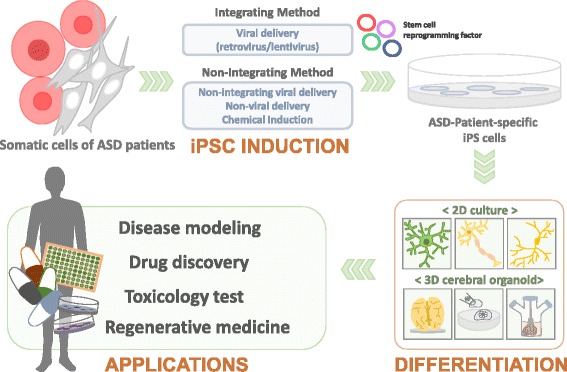
Sources of somatic cells for reprogramming
| Test methods | Description | References | |
|---|---|---|---|
| Observational report (including questionnaires) | Autism Diagnostic Observation Schedule (ADOS) | A standardized assessment in terms of objective evaluation of autistic social and communicative behavior symptoms | Lord et al. 1989 |
| Autism Diagnostic Interview-Revised (ADI-R) | An interview conducted with the parents of autistic individual to cover autistic individual’s full developmental history | Lord et al. 1994 | |
| Clinical Global Impression (CGI) Scale | A three-item scale used to assess treatment response in patients with mental disorders | Guy 1976 | |
| Childhood Autism Rating Scale (CARS) | A score calculated by subjective observation of a child’s behavior across fifteen criteria | Schopler et al. 1980; Mayes et al. 2012 | |
| Autism Behavior Checklist (ABC) | A 57-checklist of behavioral characteristics, which has been used for diagnosis of autism | Krug et al. 1980; Volkmar et al. 1988 | |
| Social Responsiveness Scale (SRS) | A quantitative measure of autistic traits completed by a caregiver familiar with the autistic individuals within 4–18 year olds | Constantino 2002; Aldridge et al. 2012 | |
| Aptitude test | Wechsler Adult Intelligence Scale (WAIS) | A form of IQ test designed to measure intelligence in adults and older adolescents, which has separate verbal and non-verbal IQ scores | Wechsler 1939 |
The most common cell type as the starting material for reprogramming process is skin dermal fibroblasts. However, since it is difficult to obtain skin biopsies from young children, especially those with autistic behavior, and the skin biopsy method using a punch is an invasive approach, it is important to obtain donor cells with high reprogramming capacity in a non-invasive way. As an alternative common cell source, peripheral blood cells are used for cellular reprogramming due to their non-invasive, easy, and routine accessibility in a clinic setting. Recent efforts in iPSC generation have attempted to develop exfoliated renal epithelial cells from urine, buccal cells, cord blood-derived cells, or keratinocytes from hair cells as cell sources for reprogramming despite concerns about purification method, culture stability for long-term passaging, reproducibility, and efficiency for reprogramming. Future advances in iPSC technology using human somatic cell types with easier access and handling, higher efficiency, and cost-effectiveness for successful reprogramming will allow development of more common customized medicines using iPSCs.
Induction methods for cellular reprogramming: integrative/non-integrative
Once somatic cells are taken from biopsies and cultured enough passages, they can be induced into stem cells using an appropriate reprogramming method. Many kinds of induction methods for cellular reprogramming have been reported depending on the delivery system of reprogramming factors or types of factors (ex, small molecules, inhibitors, etc.) as alternative reprogramming inducers. The most common method is the introduction of reprogramming factors into somatic cells via an integrating or non-integrating delivery system.
Integrating delivery system: retroviral/lentiviral vectors
Integrating methods use viral vectors such as retroviral or lentiviral vectors with high efficiency of gene delivery, although viral vectors integrate into the host cell genome (reprogramming efficiency: ~0.01–0.1 %). Generally, reprogramming factors are silenced after cellular reprogramming. However, genomic integration using viral vectors could induce reactivation of these genes, raising the possibility of oncogenesis in iPSC–derived cells or silencing of other functional genes after cellular reprogramming. Thus, many reprogramming methods without genomic integration have been described as a non-integrating approach, and some are commonly used for generation of iPSCs.
Non-integrating delivery system
| Delivery system | Pros | Cons | References | |
|---|---|---|---|---|
| Integrating method | Retrovirus | High reprogramming efficiency (~0.01–0.1 %) | Possibility of oncogenesis; silencing of functional genes | Takahashi and Yamanaka. 2006 |
| Lentivirus | High reprogramming efficiency (~0.01–0.1 %) | Possibility of oncogenesis; silencing of functional genes | Yu et al. 2007 | |
| Non-integrating method | Sendai virus | No risk of altering the host genome; high reprogramming efficiency(~1 %); easy to select iPSCs | Stringent steps to remove the reprogrammed cells of replicating virus; sensitivity of the viral RNA replicase | Fusaki et al. 2009 |
| Adenovirus | Transient, high-level expression | Low reprogramming efficiency (0.0001-0.001 %); possibility of small pieces insertion of adenoviral DNA; 3 out of 13(or approximately 23 %) were tetraploid | Stadtfeld et al. 2008 | |
| OriP/EBNA-based episomal vector | Unnecessary for viral packaging; gradual loss of cellular EV without drug selection; relatively high reprogramming efficiency of IRES2-mediated expression(~0.1 %); further addition of c-Myc and Klf4 improve the reprogramming efficiency to over 1 % | Unstable transfection efficiency | Yu et al. 2009 | |
| Piggy BAC transposons | Technical simplification (use of effortless plasmid DNA preparation and commercial transfection products); no limited range of somatic cell types for reprogramming; allow the option of xeno-free hiPSC production; accurate transgene removal through transposase expression | Labor intensive removal of multiple transposons; more CNVs in early passage than in intermediate passage; | Woltjen et al. 2009; Hussein et al. 2011 | |
| Cre-inducible/excisable lentivirus | Minimize the risk of chromosomal translocations; improve the developmental potential and differentiation capacity | Inefficient delivery of Cre; difficult to detect successful Cre-recombeniation; result in mosaic colonies; leaves 200 bp of exogenous DNA | Sommer et al. 2010; Soldner et al. 2009; Papapetrou et al. 2011 | |
| Minicircle DNA | Free of foreign or chemical elements; requiring only a single vector without the need for subsequent drug selection, vector excision, or the inclusion of oncogenes; FAD approved | Low reprogramming efficiency (~0.005 %) | Jia et al. 2010; Narsinh et al. 2011 | |
| Poly-arginine-tagged polypeptide | No risk of altering the host genome; simpler and faster approach than the genetic method | Low reprogramming efficiency (~0.006 %); requires either chemical treatment or greater than four rounds of treatment; expertise in protein chemistry and handling | Zhou et al. 2009; Kim et al. 2009 | |
| RNA-modified synthetic mRNA | Avoid the endogenous antiviral cell defense; high efficiency of over 2 %; resultant iPSC colonies emerge as early as 17 days | Labor intensive repeated transfection | Warren et al. 2010 | |
| Non-immunogenic; cost-effective; easily handled; | Relatively low and inconsistent efficiency | Hou et al. 2013 |
Generation of iPSC-derived neurons: neural differentiation
In disease modeling using patient-specific iPSCs, the most important step is to differentiate iPSCs into desired cell types with high purity. Accumulating research on vertebrate neural development has enabled us to generate specific subtypes of human neurons or glial cells from human pluripotent stem cells (PSCs) by regulating developmentally relevant signaling pathways. During embryonic development, the neural plate (embryonic neuroectoderm) is firstly specified to the forebrain, subsequently to the midbrain/hindbrain, and then to the spinal cord by caudalization signals that include retinoic acid (RA). Similarly, human PSCs can be directed to differentiate into forebrain-like neurons by inhibiting Wingless/Int proteins (Wnt) and bone morphogenic protein (BMP) signaling, midbrain/hindbrain by sonic hedgehog (SHH) and fibroblast growth factor 8 (FGF8) treatment, and spinal cord by the action of RA in vitro.
Two-dimensional neural differentiation
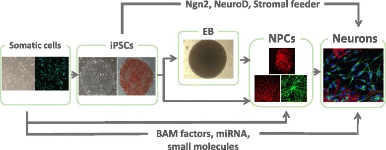
EB-dependent differentiation
| Diseases | Related genes | Neural differentiation methods | Identity of neurons | Electrophysiological proterties | Neurodevelopmental phenotypes | References |
|---|---|---|---|---|---|---|
| Rett syndrome | Methyl CpG binding protein 2 (MECP2) | Embryoid body formation | Gluramatergic & gabaergic neurons | Reduced sEPSC and sIPSC | Fewer synaptic conracts; reduced cell soma size and dendritic branching and spine density | Marchetto et al. 2010; Cheung et al. 2011; Kim et al. 2011 |
| Phelan-McDermid Syndrome (PMDS) (22q13 deletion syndrome) | Shank3 | Dual smad inhibition | Forebrain neurons | Reduced excitatory synaptic transmission | Reduced glutamatergic receptors; decreased number of synapses | Shcheglovitov et al. 2013 |
| Timothy syndrome (A member of the long QT syndromes) | CACNA1 (alpha-1 subunit of the L-type calcium channel CaV1.2) | Embryoid body formation | Cortical-enriched neuronal populations | Increase in the sustained intracellular calcium rise following membrane depolarization; wider action potentials | Decreased expression of lower corticallayers-related genes; increases in TH (tyrosine hydroxylase)-, norepinephrine- and dopamine-positice cells; activity-dependent dendrite retraction | Pasca et al. 2011; Krey et al. 2013 |
| Fragile X syndrome | Fragile X mental retardation 1 (FMR1) | Embryoid body formation | Tuj1-, MAP2- or GFAP-positive cells | Poor spontaneous synaptic activity and no glutamate reactivity | Reduced neurite numbers and neurite lengths; reduced PSD95 protein expression and reduced synaptic punctadensity; poor neuronal maturation and high gliogenic development | Sheridan et al. 2011; Telias et al. 2013 |
| Angelman syndrome | Ubiquitin protein ligase E3A (UBE3A) | Embryoid body formation | Tuj1-positive cells | Normal electrophysiological properties | Intact imprinting of UBE3A | Chamberlain et al. 2010 |
Cultivation on stromal (or mesenchymal) feeder cells
As the other neural induction method, stromal feeder-based differentiation system, which is a serum-free system without the use of either RA or EBs, has been widely used, although the molecular basis of the neural-inducing activity of stromal cells remains unclear. An initial study suggested that stromal cells induce midbrain neuronal fate by default.
Direct conversion: somatic or pluripotent stem cells to neurons/somatic cells to neural progenitor or neural stem cells
The other approach to generate human neurons is to convert human stem cells or somatic cells directly into neurons by defined specific factors or small molecules. Wernig’s group reported a simpler and direct neural conversion method from human PSCs by forced expression of only a single transcription factor, Neurogenin 2 (Ngn2), NeuroD1 or ASCL1. In human neurons induced via this method, functional synapses are rapidly formed within only 2 weeks after neural induction so that the time required to obtain mature human neurons in vitro is significantly reduced. Moreover, the most attractive point of using this method is to obtain a homogeneous cell population (~100 % of cortical neurons) differentiated from hESCs and hiPSCs. As an alternative approach to generate induced neurons, a combination of three transcription factors - BRN2 (also called Pou3f2), ASCL1 (also known as MASH1), and MYT1L (so called BAM factors) - could convert adult mouse fibroblasts directly into functional neurons without iPSC generation. The neurons generated by this method are also able to fire spontaneous action potentials and make functional synapses within as early as 2 weeks after induction in vitro. The same three transcription factors also could differentiate human stem cells and fibroblasts into neurons when combined with a transcription factor, NeuroD1, microRNAs, or small molecules. In addition, very recently, it has been reported that only small-molecule cocktails were sufficient to directly convert mouse and human fibroblasts to functional neurons without exogenous genetic factors. Likewise, rapid generation of specific subtypes of neurons directly from somatic cells makes this method an effective strategy for in vitro ASD modeling. However, a key limitation of this method is that a large number of fibroblasts might be required for reliable experiments due to their low reprogramming efficiency (at most 10–30 %), and skin biopsy cannot be conducted many times on a single patient. Therefore, in some cases, it would be desirable to convert fibroblasts into self-renewing multipotent neural progenitor cells (NPCs) or neural stem cells (NSCs), which enables us to overcome the limitations associated with low reprogramming efficiency and thereby perform high-throughput drug screening. Kim et al. described the generation of NPCs from mouse fibroblasts by transient expression of Yamanaka’s factors (Oct4, Sox2, Klf4, and c-Myc), followed by culturing in neural induction media. However, NPCs generated by this method could be expanded for only a few passages. Thier et al. have generated induced NSCs with the same classical factors (Oct4, Sox2, Klf4 and c-Myc) by strictly limiting Oct4 expression and optimizing culture conditions. In addition, forced expression of four transcription factors (BRN4/Pou3f4, SOX2, KLF4 and c-MYC) or even a single transcription factor SOX2 could also directly convert mouse or human fibroblasts into NSCs without generating a pluripotent cell state. Therefore, this direct conversion method is considered a promising method for preventing teratoma formation, which is a disadvantage of iPSCs for regenerative medicine, as well as for greatly improving low conversion efficiency from fibroblasts to neurons.
To study the pathophysiology of ASD using iPSC-derived neurons in vitro, it is important to obtain desired homogeneous neurons associated with ASD, as mentioned above. Cortical neurons have been suggested to be appropriate cell types since potential mechanisms underlying ASD include defects in cortical connectivity and neural migration to the cerebral cortex. Moreover, despite the heterogeneity of ASD, common pathways involved in synaptic development and plasticity have been proposed to be deregulated in ASD. Thus, to study developmental synaptopathy in ASD, among several protocols for neural induction, rapid generation of human cortical neurons using defined factors could be one of the best strategies for in vitro ASD modeling due to their high induction efficiency of homogenous neuronal subtype and short induction time. However, if human neurons are directly generated from stem cells or somatic cells for modeling neurodevelopmental disorders such as ASD, it might be difficult to detect developmental phenotypes during neural differentiation. Furthermore, continuous forced expression of defined factors could also mask disease phenotypes. Therefore, differentiation efficiency or stability of human neurons induced by defined factors should be improved, and comparable systematic analysis of neuronal properties such as gene expression, electrical properties, or synaptic connections in human neurons differentiated either through EB formation or by defined factors needs to be carried out.
Three-dimensional neural differentiation: cerebral organoids
Magnetic resonance imaging (MRI) studies and postmortem analysis of individual patients with ASD have consistently demonstrated anatomical abnormalities in several brain regions, which cannot be recapitulated by two-dimensional (2D) iPSC-derived neuronal culture. An iPSC-derived three-dimensional (3D) culture system termed cerebral organoid has been developed. Cerebral organoids, which develop through intrinsic self-organizing properties, can be generated from EBs grown initially in ESC medium with low bFGF and Rho kinase (ROCK) inhibitor, and they have been shown to recapitulate the complex interplay of different regions and structures of the brain. Therefore, 3D cerebral organoids derived from ASD patient-specific iPSCs would be the best in vitro model to uncover defects in cortical connectivity and neuronal migration of ASD. Indeed, Mariani et al. recently generated idiopathic ASD patient’s iPSC-derived brain organoids and showed increased production of inhibitory neurons by increased FOXG1 gene expression. However, more standardized protocols need to be developed, and further characterization and identification of neuronal cell types in specific regions of cerebral organoids should be carried out to study cortical development and for disease modeling of ASD patients.
Analyses of human iPSC-derived neurons
To use iPSC technology in modeling of various neurodevelopmental disorders including ASD in vitro, it is important to characterize disease phenotypes in disease-specific iPSC-derived neurons and validate well-known disease phenotypes to determine whether or not iPSC-derived cellular disease models could recapitulate disease phenotypes in mouse models and human patients. Because of this reason, ASD research using this technology primarily includes several studies on monogenic cases, such as Rett Syndrome, Fragile X Syndrome, and Timothy Syndrome. However, these initial studies on cellular disease phenotypes in iPSC-derived neurons from monogenic cases of ASD could be directed towards the identification of disease-relevant cellular characterization in both monogenic and idiopathic forms of ASD with high heterogeneity. In this section, we describe what phenotypic analyses of human iPSC-derived neurons can be performed to characterize and validate iPSC-derived cellular disease models.
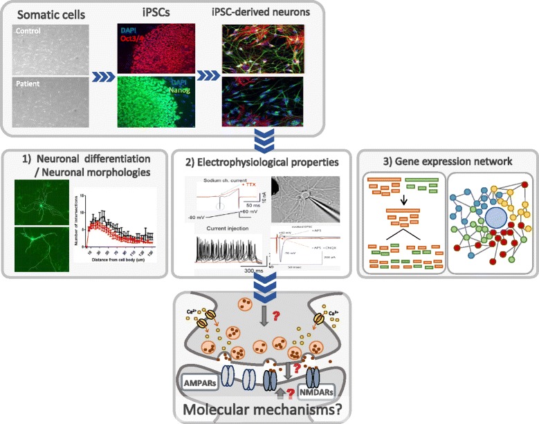
Neural differentiation and neuronal morphologies: neurite outgrowth/synapse structure
Fully differentiated neuronal cells have a distinct morphology, including distinct polarity, and extend one axon and dendritic arbors from their cell body. Therefore, the earliest phenotypes of hiPSC-derived neurons are morphological changes such as neural differentiation, neurite/axon/dendritic growth (number or length of neurite process), and synapse formation, which can be used for analysis of disease-relevant morphological phenotypic changes. During the early stages of neurogenesis, newborn neurons are NeuN-positive and PSA-NCAM-positive. These markers, together with neuronal cytoskeletal proteins Tuj1, Tau, and MAP2, can be used for measuring neural maturation efficiency or morphological changes in ASD iPSC-derived neurons. Specific neuronal gene expression as a subtype-specific marker can be also used to confirm neuronal identities. For example, glutamatergic neurons can express vGlut1 and vGlut2, GABAergic neurons express GAD65/67, and mature dopaminergic neurons express tyrosine hydroxylase (TH).
Moreover, disease phenotypes such as cortical connectivity and neural migration in ASD-derived cerebral organoids would be characterized using various markers for a specific subtype of neurons in cortical regions. In rodents, cortical glutamatergic neurons can be defined by their expression of different transcription factors. Layer 6 corticothalamic projection neurons are Tbr1-positive, layer 5 subcortical projection neurons are Ctip2-positive, layers 2–4 neurons are Cux1/2-positive, and layers 2–4 callosal projection neurons are Satb2-positive.
Electrophysiological properties
Electrophysiological characterization for basic electrical properties such as membrane potential, generation of action potentials by current injection, and synaptic properties such as appearance of spontaneous synaptic events can be applied to verify and characterize hiPSC-derived neurons. As neurons mature, resting membrane potentials (Vm) become negative (more hyperpolarized) and capacitance (Cm) increases due to increased branch numbers, leading to increased cell volume. In contrast, input resistance (Ri) decreases as channel protein expression increases. In addition, action potential-like responses induced by depolarization are resemble the mature shape of the action potential. Since neurons are able to make synapses with other neurons, synaptic properties could be also characterized as a cellular phenotype in iPSC-derived disease models. hiPSC-derived neurons from many syndromic ASD patients have defects in synaptic connectivity such as spontaneous excitatory and inhibitory currents, AMPA/NMDA current ratio, as well as intrinsic neuronal excitability (Table 3).
Gene expression network
Similar to the phenotypic analyses, transcriptional changes based on gene expression network could be characterized in ASD iPSC-derived neurons. This analysis based on a systems biology approach allows us to understand alterations of the gene network involved in neural development and functions associated with ASD. Recent studies using genome-wide weighted co-expression network analysis (GWCNA) on Timothy Syndrome (TS)-derived neural cells have shown that altered Ca2+ signaling in TS patients leads to dysregulation of calcium-dependent transcriptional regulators such as NFAT, MEF2, CREB, and FOXO as well as its downstream signals. Gene expression analysis of hiPSC-derived neurons carrying copy number variants of chromosome 15q11-q13.1 using RNA-Seq has revealed that common neuronal pathways may be disrupted in both Angelman and Dup15q syndromes.
Phenotypic analyses of ASD iPSC-derived neurons: Rett, Phelan-Mcdermid, Timothy, Fragile-X, and Angelman Syndromes
In Rett syndrome (RTT), a neurodevelopmental ASD due primarily to mutations in the methyl-CpG binding protein 2 (MECP2) gene, hiPSC-derived neural cells show reduced soma size, dendrite spine density, differentiation, and reduced spontaneous Ca2+ transient frequency in neurons and premature astroglial, as shown in human postmortem analyses. In addition, reduced frequency and amplitude of mEPSCs and mIPSCs have been observed in RTT hiPSC-derived neurons, suggesting fewer production of synapses and reduction of postsynaptic receptors. HiPSC-derived cellular disease models could be also used as a system for screening candidates for disease therapy since iPSC-derived neurons can recapitulate disease phenotypes in human and mouse models. Indeed, insulin-like growth factor 1 (IGF-1) was applied to hiPSC-derived RTT neurons and showed rescue of reduction in excitatory glutamatergic synaptic connections as in mouse models of RTT, in which reduced excitatory synaptic connections in RTT neurons could be reversed by IGF-1 application. IGF-1 is currently in clinical trials for RTT.
hiPSC-derived neurons from Phelan-Mcdermid syndrome (PMDS) patients, carrying a deletion of Shank3 protein, have significant deficits in excitatory synaptic transmission. These deficits were rescued by either wild-type Shank3 expression or IGF-1 treatment as in hiPSC-derived RTT neurons, suggesting that a common signaling pathway might be involved in the pathophysiology of ASD. Therefore, hiPSC-derived neurons could be useful as a potential drug-screening platform, as mentioned above.
Timothy syndrome (TS) is caused by a point mutation in the voltage-gated calcium channel encoded by the CACNA1 gene. TS patients iPSC-derived neurons show wider action potentials, suggesting a loss of CaV1.2 channel inactivation, abnormal expression of tyrosine hydroxylase (TH), and increased production of norepinephrine and dopamine. Activity-dependent dendritic retraction by RhoA activation independent of Ca2+ influx through CaV1.2 has also been reported in TS iPSC-derived neurons.
Neurons from Fragile-X syndrome patients-derived hiPSCs show reduced neurite numbers and lengths, poor spontaneous synaptic activity, and lack reactivity to glutamate.
In Angelman Syndrome (AS), which is caused by reduced expression of the maternal copy of the Ube3A gene in CNS, patient-derived iPSCs show retained genomic imprinting. In addition, electrophysiological recordings have detected AMPA receptor-mediated spontaneous activity in AS iPSC-derived mature neurons, suggesting that normal functional neurons can be generated from AS iPSCs.
Recently, Muotri’s group have generated an iPSC model of a nonsyndromic ASD patient carrying a de novo balanced translocation transient receptor potential channel 6 (TRPC6). TRPC6-mutant iPSC-derived neurons showed reduced neuronal arborization, fewer dendritic spines and synapses, and impaired calcium dynamics. They also found that MECP2 occupied the TRPC6 promoter region and regulated TRPC6 expression, raising the possibility of interactions among common pathways affected in nonsyndromic and syndromic ASD.
Conclusions
Perspectives: limitations and future directions
iPSC research
Despite numerous studies underlying the pathophysiological mechanism of ASD using iPSCs, several concerns should be addressed before iPSC research. Current advances in iPSC technology have allowed us to successfully derive patient-specific iPSCs regardless of their reprogramming methods. Furthermore, a recent study showed that a modular, robotic platform for iPSC reprogramming enabled automated, high-throughput conversion of skin fibroblasts into iPSCs and their characterization/differentiation with minimal manual intervention. However, it still remains unknown how to obtain qualified iPSCs and improve the quality of patient-specific iPSCs under suitable and cost-effective cultivation conditions for diverse applications, including disease modeling, drug screening, and customized therapy. As mentioned above, although iPSCs are similar to ESCs in terms of pluripotent marker gene expression, self-renewal potency, differentiation potential, and their morphology, they are not identical. Recent extensive genetic analysis using high-throughput sequencing technology or generation of single-nucleotide genome-wide maps of DNA methylation has demonstrated the genomic/epigenetic differences between iPSCs and ESCs. However, the functional consequences of their differences in vitro or in vivo are largely unknown. Moreover, iPSCs and ESCs show a wide range of clonal variations in terms of proliferation and differentiation potential. Furthermore, iPSCs derived from even the same parental somatic cells have different potential in terms of expansion or differentiation. Considerable somatic coding mutations occurring in hiPSC lines have also been reported by extensive exome analysis. The other main concern is the instability of iPSCs during passaging of clones. It has been reported that early passages of iPSCs display de novo copy number variations (CNV) during the reprogramming process. Thus, to obtain a more reliable outcome from iPSC research, generation of isogenic iPSCs using recent gene engineering technology or by establishment of at least 2–3 iPSC clones from the same parental somatic cells has been suggested. However, it remains unknown how these genetic/epigenetic alterations occur during reprogramming or expansion of iPSCs as well as how these alterations can be managed for iPSC generation or its application. Further, it remains unknown whether there is any reprogramming method to reduce or exclude these possible alterations as well as how to select the qualified iPSC clone from a variety of iPSC lines. To address these questions, further intensive works at the genetic/epigenetic/cellular levels are needed, and in vivo functional characterization of iPSC-derived cells needs to be carried out. Thus, the most important issue in iPSC generation is to establish more stable and standard protocols for safer and easier iPSC generation in diverse applications.
Although there are some differences between ESC and iPSCs, iPSCs are still the most promising choice for modeling with human cells. In mouse, iPSCs have the same potential as ESCs because a mature organism can be generated from iPSCs via blastocyst injection or tetraploid complementation. Although human iPSCs cannot be tested using these embryological methods owing to ethical issues and hiPSCs appear to be ‘primed’ PSCs as mouse Epi-stem cells, naïve human PSCs might be used as another human cellular model.
Current limitations of studies on pathophysiology using ASD iPSC-derived neurons
Besides iPSC line-to-line variations, limitations of studying ASD with hiPSC-derived neurons include phenotypic variations between neurons derived from the same iPSCs, which are based on differences between individual hiPSC-derived neurons from even a single patient due to heterogeneity of neuronal subtypes differentiated from each iPSC line even with well-defined differentiation protocols. In addition, different differentiation methods such as usage of small molecules or genes, EB formation vs. monolayer culture, concentration of small molecules and growth factors, differentiation time can also generate variations in the neuronal population. The use of cell type-specific promoters to drive expression of fluorescent markers for purification by cell sorting or identification of desired cell types would be a powerful tool to reduce variation. The surrounding environment of cells may also significantly affect the phenotypes. For example, the presence of neural progenitor cells in neuronal culture could mask disease-associated phenotypes by continuous production of newborn neurons. Therefore, to obtain reliable data using hiPSC-derived neurons from ASD patients, each experiment should be performed with multiple neuronal differentiation protocols from at least two or three independent hiPSC lines with the same mutation from multiple patients. In addition, forced expression of a transcription factor like Ngn2 would be a good method to overcome the above described issues, in which almost ~100 % of cortical neurons at a similar maturation stage could be generated, and the neurons showed their synaptic phenotypes as early as 3 weeks after forced Ngn2 expression.
As mentioned above, widespread genetic variations could exist between iPSC lines themselves derived from unrelated individuals. Therefore, genetically related family member-derived control lines could possibly be used to reduce variability of phenotypes, although it would not completely remove the possibility that even a single genetic difference could potentially affect observable phenotypes. Another possible way is to use gene correction methods since the ideal controls would be those that have the same genetic background except only the specific genetic defect found in the patient. Many well-known syndromic ASD-related genetic variants can be modeled with “isogenic” cell lines, where a patient-derived iPSC line could be gene-corrected using ZFNs, TALENs, or CRISPR-Cas9 technologies, reverting a mutant line to wild-type or vice versa.
As iPSCs are an in vitro culture system, they lack many characteristics of a developing and mature brain physiology in vivo. Therefore, it is difficult to study neuronal circuitry and organization using iPSC-derived neurons under 2D conditions, particularly when investigating phenotypes unique to specific neuronal circuits of the adult brain. One way to avoid these problems is to xenograft iPSC-derived neural progenitor cells (NPCs) into embryonic rodent brains to allow them to integrate into developing neural networks and mature in vivo. Cerebral “organoids” is another possible way to study disease phenotypes in a specific cell type or group of cell fates in the context of 3D model of human neurodevelopment, as reported by Mariani et al..
In summary, we can generate hiPSC-derived neurons from fibroblasts and other somatic cells of ASD patients to investigate alterations of neuronal connectivity, synaptic maturation, and functions. In addition, direct conversion of fibroblasts from ASD patients into neurons or NPC/NSCs would be used as an alternative in vitro model of ASD in the near future. However, we need to realize that hiPSC-based studies of ASD pathophysiology will not completely replace human postmortem and mouse genetic studies. Nevertheless, disease modeling with hiPSC-derived neurons combined with their comprehensive molecular and functional characterization will be a new and strong tool for understanding complex neurodevelopmental disorder, ASD.
Sources
- Baird G, Simonoff E, Pickles A, Chandler S, Loucas T, Meldrum DPrevalence of disorders of the autism spectrum in a population cohort of children in South Thames: the Special Needs and Autism Project (SNAP)Lancet2006368210215 https://doi.org/10.1016/S0140-6736(06)69041-7
- Kogan MD, Blumberg SJ, Schieve LA, Boyle CA, Perrin JM, Ghandour RMPrevalence of parent-reported diagnosis of autism spectrum disorder among children in the US, 2007Pediatrics200912413951403 https://doi.org/10.1542/peds.2009-1522
- Pinto D, Delaby E, Merico D, Barbosa M, Merikangas A, Klei LConvergence of genes and cellular pathways dysregulated in autism spectrum disordersAm J Hum Genet201494677694 https://doi.org/10.1016/j.ajhg.2014.03.018
- Jeremy Willsey A, State MWAutism spectrum disorders: from genes to neurobiologyCurr Opin Neurobiol201530C9299 https://doi.org/10.1016/j.conb.2014.10.015
- Devlin B, Scherer SWGenetic architecture in autism spectrum disorderCurr Opin Genet Dev201222229237 https://doi.org/10.1016/j.gde.2012.03.002
- Zoghbi HY, Bear MFSynaptic dysfunction in neurodevelopmental disorders associated with autism and intellectual disabilitiesCold Spring Harb Perspect Biol201243 https://doi.org/10.1101/cshperspect.a009886
- An JY, Cristino AS, Zhao Q, Edson J, Williams SM, Ravine DTowards a molecular characterization of autism spectrum disorders: an exome sequencing and systems approachTransl Psychiatry20144e394 https://doi.org/10.1038/tp.2014.38
- Ebrahimi-Fakhari D, Sahin MAutism and the synapse: emerging mechanisms and mechanism-based therapiesCurr Opin Neurol20152891102 https://doi.org/10.1097/WCO.0000000000000186
- Bourgeron TFrom the genetic architecture to synaptic plasticity in autism spectrum disorderNat Rev Neurosci201516551563 https://doi.org/10.1038/nrn3992
- Won H, Lee HR, Gee HY, Mah W, Kim JI, Lee JAutistic-like social behaviour in Shank2-mutant mice improved by restoring NMDA receptor functionNature2012486261265 https://doi.org/10.1038/nature11208
- Banerjee S, Riordan M, Bhat MAGenetic aspects of autism spectrum disorders: insights from animal modelsFront Cell Neurosci2014858 https://doi.org/10.3389/fncel.2014.00058
- Moy SS, Nadler JJAdvances in behavioral genetics: mouse models of autismMol Psychiatry200813426 https://doi.org/10.1038/sj.mp.4002082
- Ey E, Leblond CS, Bourgeron TBehavioral profiles of mouse models for autism spectrum disordersAutism Res20114516 https://doi.org/10.1002/aur.175
- Tordjman S, Drapier D, Bonnot O, Graignic R, Fortes S, Cohen DAnimal models relevant to schizophrenia and autism: validity and limitationsBehav Genet2007376178 https://doi.org/10.1007/s10519-006-9120-5
- Tania M, Khan MA, Xia KRecent advances in animal model experimentation in autism researchActa Neuropsychiatr201426264271 https://doi.org/10.1017/neu.2013.58
- Thomson JA, Itskovitz-Eldor J, Shapiro SS, Waknitz MA, Swiergiel JJ, Marshall VSEmbryonic stem cell lines derived from human blastocystsScience (New York, NY)199828211451147 https://doi.org/10.1126/science.282.5391.1145
- Shi Y, Kirwan P, Smith J, Robinson HP, Livesey FJHuman cerebral cortex development from pluripotent stem cells to functional excitatory synapsesNat Neurosci201215477–486S471 https://doi.org/10.1038/nn.3041
- Liu EY, Scott CTGreat expectations: autism spectrum disorder and induced pluripotent stem cell technologiesStem Cell Rev201410145150 https://doi.org/10.1007/s12015-014-9497-0
- Kim DS, Ross PJ, Zaslavsky K, Ellis JOptimizing neuronal differentiation from induced pluripotent stem cells to model ASDFront Cell Neurosci20148109 https://doi.org/10.3389/fncel.2014.00109
- Prilutsky D, Palmer NP, Smedemark-Margulies N, Schlaeger TM, Margulies DM, Kohane ISiPSC-derived neurons as a higher-throughput readout for autism: promises and pitfallsTrends Mol Med20142091104 https://doi.org/10.1016/j.molmed.2013.11.004
- Ding Q, Lee YK, Schaefer EA, Peters DT, Veres A, Kim KA TALEN genome-editing system for generating human stem cell-based disease modelsCell Stem Cell201312238251 https://doi.org/10.1016/j.stem.2012.11.011
- Lombardo A, Genovese P, Beausejour CM, Colleoni S, Lee YL, Kim KAGene editing in human stem cells using zinc finger nucleases and integrase-defective lentiviral vector deliveryNat Biotechnol20072512981306 https://doi.org/10.1038/nbt1353
- Kim Y, Kweon J, Kim A, Chon JK, Yoo JY, Kim HJA library of TAL effector nucleases spanning the human genomeNat Biotechnol201331251258 https://doi.org/10.1038/nbt.2517
- Smith C, Abalde-Atristain L, He C, Brodsky BR, Braunstein EM, Chaudhari PEfficient and allele-specific genome editing of disease loci in human iPSCsMolecular therapy: the journal of the American Society of Gene Therapy201523570577 https://doi.org/10.1038/mt.2014.226
- Chiba K, Hockemeyer DGenome editing in human pluripotent stem cells using site-specific nucleasesMethods in molecular biology (Clifton, NJ)20151239267280 https://doi.org/10.1007/978-1-4939-1862-1_15
- Okano H, Yamanaka SiPS cell technologies: significance and applications to CNS regeneration and diseaseMol Brain2014722 https://doi.org/10.1186/1756-6606-7-22
- Velasco I, Salazar P, Giorgetti A, Ramos-Mejia V, Castano J, Romero-Moya DConcise review: generation of neurons from somatic cells of healthy individuals and neurological patients through induced pluripotency or direct conversionStem cells (Dayton, Ohio)20143228112817 https://doi.org/10.1002/stem.1782
- Xu J, Du Y, Deng HDirect Lineage Reprogramming: Strategies, Mechanisms, and ApplicationsCell Stem Cell201516119134 https://doi.org/10.1016/j.stem.2015.01.013
- Kim J, Ambasudhan R, Ding SDirect lineage reprogramming to neural cellsCurr Opin Neurobiol201222778784 https://doi.org/10.1016/j.conb.2012.05.001
- Okano H, Nakamura M, Yoshida K, Okada Y, Tsuji O, Nori SSteps toward safe cell therapy using induced pluripotent stem cellsCirc Res2013112523533 https://doi.org/10.1161/CIRCRESAHA.111.256149
- Takahashi K, Tanabe K, Ohnuki M, Narita M, Ichisaka T, Tomoda KInduction of pluripotent stem cells from adult human fibroblasts by defined factorsCell2007131861872 https://doi.org/10.1016/j.cell.2007.11.019
- Takahashi K, Yamanaka SInduction of pluripotent stem cells from mouse embryonic and adult fibroblast cultures by defined factorsCell2006126663676 https://doi.org/10.1016/j.cell.2006.07.024
- Singh VK, Kalsan M, Kumar N, Saini A, Chandra RInduced pluripotent stem cells: applications in regenerative medicine, disease modeling, and drug discoveryFront Cell Dev Biol201532 https://doi.org/10.3389/fcell.2015.00002
- Rony IK, Baten A, Bloomfield JA, Islam ME, Billah MM, Islam KDInducing pluripotency in vitro: recent advances and highlights in induced pluripotent stem cells generation and pluripotency reprogrammingCell Prolif201548140156 https://doi.org/10.1111/cpr.12162
- Li J, Song W, Pan G, Zhou JAdvances in understanding the cell types and approaches used for generating induced pluripotent stem cellsJ Hematol Oncol2014750 https://doi.org/10.1186/s13045-014-0050-z
- Zhou YY, Zeng FIntegration-free methods for generating induced pluripotent stem cellsGenomics Proteomics Bioinformatics201311284287 https://doi.org/10.1016/j.gpb.2013.09.008
- Petit I, Kesner NS, Karry R, Robicsek O, Aberdam E, Muller FJInduced pluripotent stem cells from hair follicles as a cellular model for neurodevelopmental disordersStem Cell Res20128134140 https://doi.org/10.1016/j.scr.2011.09.003
- Raab S, Klingenstein M, Liebau S, Linta LA Comparative View on Human Somatic Cell Sources for iPSC GenerationStem Cells Int20142014768391 https://doi.org/10.1155/2014/768391
- Lord C, Rutter M, Goode S, Heemsbergen J, Jordan H, Mawhood LAutism diagnostic observation schedule: a standardized observation of communicative and social behaviorJ Autism Dev Disord198919185212 https://doi.org/10.1007/BF02211841
- Lord C, Rutter M, Le Couteur AAutism Diagnostic Interview-Revised: a revised version of a diagnostic interview for caregivers of individuals with possible pervasive developmental disordersJ Autism Dev Disord199424659685 https://doi.org/10.1007/BF02172145
- Guy WECDEU Assessment Manual for Psychopharmacology1976Rockville, MdUS Department of Health, Education, and Welfare
- Schopler E, Reichler RJ, DeVellis RF, Daly KToward objective classification of childhood autism: Childhood Autism Rating Scale (CARS)J Autism Dev Disord19801091103 https://doi.org/10.1007/BF02408436
- Mayes SD, Calhoun SL, Murray MJ, Morrow JD, Yurich KKL, Cothren SUse of the Childhood Autism Rating Scale (CARS) for Children With High Functioning Autism or Asperger SyndromeFocus on Autism and Other Developmental Disabilities2012273138 https://doi.org/10.1177/1088357611406902
- Krug DA, Arick J, Almond PBehavior checklist for identifying severely handicapped individuals with high levels of autistic behaviorJ Child Psychol Psychiatry198021221229 https://doi.org/10.1111/j.1469-7610.1980.tb01797.x
- Volkmar FR, Cicchetti DV, Dykens E, Sparrow SS, Leckman JF, Cohen DJAn evaluation of the Autism Behavior ChecklistJ Autism Dev Disord1988188197 https://doi.org/10.1007/BF02211820
- Constantino JThe social responsiveness scale2002Los AngelesWestern Psychological Services
- Aldridge FJ, Gibbs VM, Schmidhofer K, Williams MInvestigating the clinical usefulness of the Social Responsiveness Scale (SRS) in a tertiary level, autism spectrum disorder specific assessment clinicJ Autism Dev Disord201242294300 https://doi.org/10.1007/s10803-011-1242-9
- Wechsler DThe Measurement of Adult Intelligence1939BaltimoreWilliams & Wilkins
- Dowey SN, Huang X, Chou BK, Ye Z, Cheng LGeneration of integration-free human induced pluripotent stem cells from postnatal blood mononuclear cells by plasmid vector expressionNat Protoc2012720132021 https://doi.org/10.1038/nprot.2012.121
- Quintana-Bustamante O, Segovia JCGeneration of Patient-Specific induced Pluripotent Stem Cell from Peripheral Blood Mononuclear Cells by Sendai Reprogramming Vectors2014Clifton, N.JMethods in molecular biology https://doi.org/10.1007/7651_2014_170
- Yu J, Vodyanik MA, Smuga-Otto K, Antosiewicz-Bourget J, Frane JL, Tian SInduced pluripotent stem cell lines derived from human somatic cellsScience (New York, NY)200731819171920 https://doi.org/10.1126/science.1151526
- Maherali N, Hochedlinger KGuidelines and techniques for the generation of induced pluripotent stem cellsCell Stem Cell20083595605 https://doi.org/10.1016/j.stem.2008.11.008
- Nori S, Okada Y, Nishimura S, Sasaki T, Itakura G, Kobayashi YLong-term safety issues of iPSC-based cell therapy in a spinal cord injury model: oncogenic transformation with epithelial-mesenchymal transitionStem Cell Rep20154360373 https://doi.org/10.1016/j.stemcr.2015.01.006
- Choi HW, Kim JS, Choi S, Hong YJ, Kim MJ, Seo HGNeural stem cells differentiated from iPS cells spontaneously regain pluripotencyStem cells (Dayton, Ohio)20143225962604 https://doi.org/10.1002/stem.1757
- Fusaki N, Ban H, Nishiyama A, Saeki K, Hasegawa MEfficient induction of transgene-free human pluripotent stem cells using a vector based on Sendai virus, an RNA virus that does not integrate into the host genomeProc Jpn Acad Ser B Phys Biol Sci200985348362 https://doi.org/10.2183/pjab.85.348
- Stadtfeld M, Nagaya M, Utikal J, Weir G, Hochedlinger KInduced pluripotent stem cells generated without viral integrationScience (New York, NY)2008322945949 https://doi.org/10.1126/science.1162494
- Okita K, Nakagawa M, Hyenjong H, Ichisaka T, Yamanaka SGeneration of mouse induced pluripotent stem cells without viral vectorsScience (New York, NY)2008322949953 https://doi.org/10.1126/science.1164270
- Somers A, Jean JC, Sommer CA, Omari A, Ford CC, Mills JAGeneration of transgene-free lung disease-specific human induced pluripotent stem cells using a single excisable lentiviral stem cell cassetteStem cells (Dayton, Ohio)20102817281740 https://doi.org/10.1002/stem.495
- Woltjen K, Michael IP, Mohseni P, Desai R, Mileikovsky M, Hamalainen RpiggyBac transposition reprograms fibroblasts to induced pluripotent stem cellsNature2009458766770 https://doi.org/10.1038/nature07863
- Kim D, Kim CH, Moon JI, Chung YG, Chang MY, Han BSGeneration of human induced pluripotent stem cells by direct delivery of reprogramming proteinsCell Stem Cell20094472476 https://doi.org/10.1016/j.stem.2009.05.005
- Warren L, Manos PD, Ahfeldt T, Loh YH, Li H, Lau FHighly efficient reprogramming to pluripotency and directed differentiation of human cells with synthetic modified mRNACell Stem Cell20107618630 https://doi.org/10.1016/j.stem.2010.08.012
- Dang J, Rana TMEnhancing Induced Pluripotent Stem Cell Generation by MicroRNA2015Clifton, N.JMethods in molecular biology https://doi.org/10.1007/7651_2015_201
- Yoshioka N, Gros E, Li HR, Kumar S, Deacon DC, Maron CEfficient generation of human iPSCs by a synthetic self-replicative RNACell Stem Cell201313246254 https://doi.org/10.1016/j.stem.2013.06.001
- Woodard LE, Wilson MHpiggyBac-ing models and new therapeutic strategiesTrends Biotechnol201533525533 https://doi.org/10.1016/j.tibtech.2015.06.009
- Lin T, Wu SReprogramming with Small Molecules instead of Exogenous Transcription FactorsStem Cells Int20152015794632 https://doi.org/10.1155/2015/794632
- Ban H, Nishishita N, Fusaki N, Tabata T, Saeki K, Shikamura MEfficient generation of transgene-free human induced pluripotent stem cells (iPSCs) by temperature-sensitive Sendai virus vectorsProc Natl Acad Sci U S A20111081423414239 https://doi.org/10.1073/pnas.1103509108
- Fujie Y, Fusaki N, Katayama T, Hamasaki M, Soejima Y, Soga MNew type of Sendai virus vector provides transgene-free iPS cells derived from chimpanzee bloodPLoS One20149e113052 https://doi.org/10.1371/journal.pone.0113052
- Zhou W, Freed CRAdenoviral gene delivery can reprogram human fibroblasts to induced pluripotent stem cellsStem cells (Dayton, Ohio)20092726672674 https://doi.org/10.1002/stem.201
- Okita K, Matsumura Y, Sato Y, Okada A, Morizane A, Okamoto SA more efficient method to generate integration-free human iPS cellsNat Methods20118409412 https://doi.org/10.1038/nmeth.1591
- Kaji K, Norrby K, Paca A, Mileikovsky M, Mohseni P, Woltjen KVirus-free induction of pluripotency and subsequent excision of reprogramming factorsNature2009458771775 https://doi.org/10.1038/nature07864
- Chang CW, Lai YS, Pawlik KM, Liu K, Sun CW, Li CPolycistronic lentiviral vector for “hit and run” reprogramming of adult skin fibroblasts to induced pluripotent stem cellsStem cells (Dayton, Ohio)20092710421049 https://doi.org/10.1002/stem.39
- Ren X, Tahimic CG, Katoh M, Kurimasa A, Inoue T, Oshimura MHuman artificial chromosome vectors meet stem cells: new prospects for gene deliveryStem Cell Rev200624350 https://doi.org/10.1007/s12015-006-0008-9
- Jia F, Wilson KD, Sun N, Gupta DM, Huang M, Li ZA nonviral minicircle vector for deriving human iPS cellsNat Methods20107197199 https://doi.org/10.1038/nmeth.1426
- Hiratsuka M, Uno N, Ueda K, Kurosaki H, Imaoka N, Kazuki KIntegration-free iPS cells engineered using human artificial chromosome vectorsPLoS One20116e25961 https://doi.org/10.1371/journal.pone.0025961
- Narsinh KH, Jia F, Robbins RC, Kay MA, Longaker MT, Wu JCGeneration of adult human induced pluripotent stem cells using nonviral minicircle DNA vectorsNat Protoc201167888 https://doi.org/10.1038/nprot.2010.173
- Malik N, Rao MSA review of the methods for human iPSC derivationMethods in molecular biology (Clifton, NJ)20139972333 https://doi.org/10.1007/978-1-62703-348-0_3
- Huangfu D, Maehr R, Guo W, Eijkelenboom A, Snitow M, Chen AEInduction of pluripotent stem cells by defined factors is greatly improved by small-molecule compoundsNat Biotechnol200826795797 https://doi.org/10.1038/nbt1418
- Huangfu D, Osafune K, Maehr R, Guo W, Eijkelenboom A, Chen SInduction of pluripotent stem cells from primary human fibroblasts with only Oct4 and Sox2Nat Biotechnol20082612691275 https://doi.org/10.1038/nbt.1502
- Li W, Zhou H, Abujarour R, Zhu S, Young Joo J, Lin TGeneration of human-induced pluripotent stem cells in the absence of exogenous Sox2Stem cells (Dayton, Ohio)20092729923000 https://doi.org/10.1002/stem.240
- Hou P, Li Y, Zhang X, Liu C, Guan J, Li HPluripotent stem cells induced from mouse somatic cells by small-molecule compoundsScience (New York, NY)2013341651654 https://doi.org/10.1126/science.1239278
- Schlaeger TM, Daheron L, Brickler TR, Entwisle S, Chan K, Cianci AA comparison of non-integrating reprogramming methodsNat Biotechnol2015335863 https://doi.org/10.1038/nbt.3070
- Gaspard N, Vanderhaeghen PMechanisms of neural specification from embryonic stem cellsCurr Opin Neurobiol2010203743 https://doi.org/10.1016/j.conb.2009.12.001
- Perrier AL, Tabar V, Barberi T, Rubio ME, Bruses J, Topf NDerivation of midbrain dopamine neurons from human embryonic stem cellsProc Natl Acad Sci U S A20041011254312548 https://doi.org/10.1073/pnas.0404700101
- Lee SH, Lumelsky N, Studer L, Auerbach JM, McKay RDEfficient generation of midbrain and hindbrain neurons from mouse embryonic stem cellsNat Biotechnol200018675679 https://doi.org/10.1038/76536
- Wichterle H, Lieberam I, Porter JA, Jessell TMDirected differentiation of embryonic stem cells into motor neuronsCell2002110385397 https://doi.org/10.1016/S0092-8674(02)00835-8
- Li XJ, Du ZW, Zarnowska ED, Pankratz M, Hansen LO, Pearce RASpecification of motoneurons from human embryonic stem cellsNat Biotechnol200523215221 https://doi.org/10.1038/nbt1063
- Zhang SC, Wernig M, Duncan ID, Brustle O, Thomson JAIn vitro differentiation of transplantable neural precursors from human embryonic stem cellsNat Biotechnol20011911291133 https://doi.org/10.1038/nbt1201-1129
- Okabe S, Forsberg-Nilsson K, Spiro AC, Segal M, McKay RDDevelopment of neuronal precursor cells and functional postmitotic neurons from embryonic stem cells in vitroMech Dev19965989102 https://doi.org/10.1016/0925-4773(96)00572-2
- Imaizumi Y, Okada Y, Akamatsu W, Koike M, Kuzumaki N, Hayakawa HMitochondrial dysfunction associated with increased oxidative stress and alpha-synuclein accumulation in PARK2 iPSC-derived neurons and postmortem brain tissueMol Brain2012535 https://doi.org/10.1186/1756-6606-5-35
- Numasawa-Kuroiwa Y, Okada Y, Shibata S, Kishi N, Akamatsu W, Shoji MInvolvement of ER stress in dysmyelination of Pelizaeus-Merzbacher Disease with PLP1 missense mutations shown by iPSC-derived oligodendrocytesStem Cell Rep20142648661 https://doi.org/10.1016/j.stemcr.2014.03.007
- Barberi T, Klivenyi P, Calingasan NY, Lee H, Kawamata H, Loonam KNeural subtype specification of fertilization and nuclear transfer embryonic stem cells and application in parkinsonian miceNat Biotechnol20032112001207 https://doi.org/10.1038/nbt870
- Kawasaki H, Mizuseki K, Nishikawa S, Kaneko S, Kuwana Y, Nakanishi SInduction of midbrain dopaminergic neurons from ES cells by stromal cell-derived inducing activityNeuron2000283140 https://doi.org/10.1016/S0896-6273(00)00083-0
- Vierbuchen T, Ostermeier A, Pang ZP, Kokubu Y, Sudhof TC, Wernig MDirect conversion of fibroblasts to functional neurons by defined factorsNature201046310351041 https://doi.org/10.1038/nature08797
- Pang ZP, Yang N, Vierbuchen T, Ostermeier A, Fuentes DR, Yang TQInduction of human neuronal cells by defined transcription factorsNature2011476220223 https://doi.org/10.1038/nature10202
- Ambasudhan R, Talantova M, Coleman R, Yuan X, Zhu S, Lipton SADirect reprogramming of adult human fibroblasts to functional neurons under defined conditionsCell Stem Cell20119113118 https://doi.org/10.1016/j.stem.2011.07.002
- Yoo AS, Sun AX, Li L, Shcheglovitov A, Portmann T, Li YMicroRNA-mediated conversion of human fibroblasts to neuronsNature2011476228231 https://doi.org/10.1038/nature10323
- Zhang Y, Pak C, Han Y, Ahlenius H, Zhang Z, Chanda SRapid single-step induction of functional neurons from human pluripotent stem cellsNeuron201378785798 https://doi.org/10.1016/j.neuron.2013.05.029
- Babos K, Ichida JKSmall Molecules Take a Big Step by Converting Fibroblasts into NeuronsCell Stem Cell201517127129 https://doi.org/10.1016/j.stem.2015.07.018
- Li X, Zuo X, Jing J, Ma Y, Wang J, Liu DSmall-Molecule-Driven Direct Reprogramming of Mouse Fibroblasts into Functional NeuronsCell Stem Cell201517195203 https://doi.org/10.1016/j.stem.2015.06.003
- Hu W, Qiu B, Guan W, Wang Q, Wang M, Li WDirect Conversion of Normal and Alzheimer’s Disease Human Fibroblasts into Neuronal Cells by Small MoleculesCell Stem Cell201517204212 https://doi.org/10.1016/j.stem.2015.07.006
- Nguyen HN, Byers B, Cord B, Shcheglovitov A, Byrne J, Gujar PLRRK2 mutant iPSC-derived DA neurons demonstrate increased susceptibility to oxidative stressCell Stem Cell20118267280 https://doi.org/10.1016/j.stem.2011.01.013
- Seibler P, Graziotto J, Jeong H, Simunovic F, Klein C, Krainc DMitochondrial Parkin recruitment is impaired in neurons derived from mutant PINK1 induced pluripotent stem cellsThe Journal of neuroscience: the official journal of the Society for Neuroscience20113159705976 https://doi.org/10.1523/JNEUROSCI.4441-10.2011
- Bain G, Kitchens D, Yao M, Huettner JE, Gottlieb DIEmbryonic stem cells express neuronal properties in vitroDev Biol1995168342357 https://doi.org/10.1006/dbio.1995.1085
- Marchetto MC, Carromeu C, Acab A, Yu D, Yeo GW, Mu YA model for neural development and treatment of Rett syndrome using human induced pluripotent stem cellsCell2010143527539 https://doi.org/10.1016/j.cell.2010.10.016
- Cheung AY, Horvath LM, Grafodatskaya D, Pasceri P, Weksberg R, Hotta AIsolation of MECP2-null Rett Syndrome patient hiPS cells and isogenic controls through X-chromosome inactivationHum Mol Genet20112021032115 https://doi.org/10.1093/hmg/ddr093
- Kim KY, Hysolli E, Park IHNeuronal maturation defect in induced pluripotent stem cells from patients with Rett syndromeProc Natl Acad Sci U S A20111081416914174 https://doi.org/10.1073/pnas.1018979108
- Pasca SP, Portmann T, Voineagu I, Yazawa M, Shcheglovitov A, Pasca AMUsing iPSC-derived neurons to uncover cellular phenotypes associated with Timothy syndromeNat Med20111716571662 https://doi.org/10.1038/nm.2576
- Krey JF, Pasca SP, Shcheglovitov A, Yazawa M, Schwemberger R, Rasmusson RTimothy syndrome is associated with activity-dependent dendritic retraction in rodent and human neuronsNat Neurosci201316201209 https://doi.org/10.1038/nn.3307
- Sheridan SD, Theriault KM, Reis SA, Zhou F, Madison JM, Daheron LEpigenetic characterization of the FMR1 gene and aberrant neurodevelopment in human induced pluripotent stem cell models of fragile X syndromePLoS One20116e26203 https://doi.org/10.1371/journal.pone.0026203
- Telias M, Segal M, Ben-Yosef DNeural differentiation of Fragile X human Embryonic Stem Cells reveals abnormal patterns of development despite successful neurogenesisDev Biol20133743245 https://doi.org/10.1016/j.ydbio.2012.11.031
- Chamberlain SJ, Chen PF, Ng KY, Bourgois-Rocha F, Lemtiri-Chlieh F, Levine ESInduced pluripotent stem cell models of the genomic imprinting disorders Angelman and Prader-Willi syndromesProc Natl Acad Sci U S A20101071766817673 https://doi.org/10.1073/pnas.1004487107
- Sandoe J, Eggan KOpportunities and challenges of pluripotent stem cell neurodegenerative disease modelsNat Neurosci201316780789 https://doi.org/10.1038/nn.3425
- Han SS, Williams LA, Eggan KCConstructing and deconstructing stem cell models of neurological diseaseNeuron201170626644 https://doi.org/10.1016/j.neuron.2011.05.003
- Chambers SM, Fasano CA, Papapetrou EP, Tomishima M, Sadelain M, Studer LHighly efficient neural conversion of human ES and iPS cells by dual inhibition of SMAD signalingNat Biotechnol200927275280 https://doi.org/10.1038/nbt.1529
- Chambers SM, Mica Y, Lee G, Studer L, Tomishima MJDual-SMAD Inhibition/WNT Activation-Based Methods to Induce Neural Crest and Derivatives from Human Pluripotent Stem Cells2013Clifton, N.JMethods in molecular biology https://doi.org/10.1007/7651_2013_59
- Pera MF, Andrade J, Houssami S, Reubinoff B, Trounson A, Stanley EGRegulation of human embryonic stem cell differentiation by BMP-2 and its antagonist nogginJ Cell Sci200411712691280 https://doi.org/10.1242/jcs.00970
- Shcheglovitov A, Shcheglovitova O, Yazawa M, Portmann T, Shu R, Sebastiano VSHANK3 and IGF1 restore synaptic deficits in neurons from 22q13 deletion syndrome patientsNature2013503267271 https://doi.org/10.1038/nature12618
- Chanda S, Ang CE, Davila J, Pak C, Mall M, Lee QYGeneration of induced neuronal cells by the single reprogramming factor ASCL1Stem Cell Rep20143282296 https://doi.org/10.1016/j.stemcr.2014.05.020
- Chanda S, Marro S, Wernig M, Sudhof TCNeurons generated by direct conversion of fibroblasts reproduce synaptic phenotype caused by autism-associated neuroligin-3 mutationProc Natl Acad Sci U S A20131101662216627 https://doi.org/10.1073/pnas.1316240110
- Yoo AS, Sun AX, Li L, Shcheglovitov A, Portmann T, Li YMicroRNA-mediated conversion of human fibroblasts to neuronsNature2011476228231 https://doi.org/10.1038/nature10323
- Ladewig J, Mertens J, Kesavan J, Doerr J, Poppe D, Glaue FSmall molecules enable highly efficient neuronal conversion of human fibroblastsNat Methods20129575578 https://doi.org/10.1038/nmeth.1972
- Kim J, Efe JA, Zhu S, Talantova M, Yuan X, Wang SDirect reprogramming of mouse fibroblasts to neural progenitorsProc Natl Acad Sci U S A201110878387843 https://doi.org/10.1073/pnas.1103113108
- Thier M, Worsdorfer P, Lakes YB, Gorris R, Herms S, Opitz TDirect conversion of fibroblasts into stably expandable neural stem cellsCell Stem Cell201210473479 https://doi.org/10.1016/j.stem.2012.03.003
- Han DW, Tapia N, Hermann A, Hemmer K, Hoing S, Arauzo-Bravo MJDirect reprogramming of fibroblasts into neural stem cells by defined factorsCell Stem Cell201210465472 https://doi.org/10.1016/j.stem.2012.02.021
- Ring KL, Tong LM, Balestra ME, Javier R, Andrews-Zwilling Y, Li GDirect reprogramming of mouse and human fibroblasts into multipotent neural stem cells with a single factorCell Stem Cell201211100109 https://doi.org/10.1016/j.stem.2012.05.018
- Kim SM, Flasskamp H, Hermann A, Arauzo-Bravo MJ, Lee SC, Lee SHDirect conversion of mouse fibroblasts into induced neural stem cellsNat Protoc20149871881 https://doi.org/10.1038/nprot.2014.056
- Just MA, Cherkassky VL, Keller TA, Kana RK, Minshew NJFunctional and anatomical cortical underconnectivity in autism: evidence from an FMRI study of an executive function task and corpus callosum morphometryCerebral cortex (New York, NY: 1991)200717951961 https://doi.org/10.1093/cercor/bhl006
- Brennand KJ, Gage FHModeling psychiatric disorders through reprogrammingDis Model Mech201252632 https://doi.org/10.1242/dmm.008268
- Amaral DG, Schumann CM, Nordahl CWNeuroanatomy of autismTrends Neurosci200831137145 https://doi.org/10.1016/j.tins.2007.12.005
- Lancaster MA, Renner M, Martin CA, Wenzel D, Bicknell LS, Hurles MECerebral organoids model human brain development and microcephalyNature2013501373379 https://doi.org/10.1038/nature12517
- Lancaster MA, Knoblich JAGeneration of cerebral organoids from human pluripotent stem cellsNat Protoc2014923292340 https://doi.org/10.1038/nprot.2014.158
- Mariani J, Coppola G, Zhang P, Abyzov A, Provini L, Tomasini LFOXG1-Dependent Dysregulation of GABA/Glutamate Neuron Differentiation in Autism Spectrum DisordersCell2015162375390 https://doi.org/10.1016/j.cell.2015.06.034
- Farra N, Zhang WB, Pasceri P, Eubanks JH, Salter MW, Ellis JRett syndrome induced pluripotent stem cell-derived neurons reveal novel neurophysiological alterationsMol Psychiatry20121712611271 https://doi.org/10.1038/mp.2011.180
- Urbach A, Bar-Nur O, Daley GQ, Benvenisty NDifferential modeling of fragile X syndrome by human embryonic stem cells and induced pluripotent stem cellsCell Stem Cell20106407411 https://doi.org/10.1016/j.stem.2010.04.005
- Mullen RJ, Buck CR, Smith AMNeuN, a neuronal specific nuclear protein in vertebratesDevelopment1992116201211 https://doi.org/10.1242/dev.116.1.201
- Bonfanti L, Theodosis DTExpression of polysialylated neural cell adhesion molecule by proliferating cells in the subependymal layer of the adult rat, in its rostral extension and in the olfactory bulbNeuroscience199462291305 https://doi.org/10.1016/0306-4522(94)90333-6
- von Bohlen Und Halbach OImmunohistological markers for staging neurogenesis in adult hippocampusCell Tissue Res2007329409420 https://doi.org/10.1007/s00441-007-0432-4
- Takamori S, Rhee JS, Rosenmund C, Jahn RIdentification of a vesicular glutamate transporter that defines a glutamatergic phenotype in neuronsNature2000407189194 https://doi.org/10.1038/35025070
- Kaufman DL, Houser CR, Tobin AJTwo forms of the gamma-aminobutyric acid synthetic enzyme glutamate decarboxylase have distinct intraneuronal distributions and cofactor interactionsJ Neurochem199156720723 https://doi.org/10.1111/j.1471-4159.1991.tb08211.x
- Pickel VM, Joh TH, Field PM, Becker CG, Reis DJCellular localization of tyrosine hydroxylase by immunohistochemistryThe journal of histochemistry and cytochemistry: official journal of the Histochemistry Society197523112 https://doi.org/10.1177/23.1.234988
- Frantz GD, Weimann JM, Levin ME, McConnell SKOtx1 and Otx2 define layers and regions in developing cerebral cortex and cerebellumThe Journal of neuroscience: the official journal of the Society for Neuroscience19941457255740 https://doi.org/10.1523/JNEUROSCI.14-10-05725.1994
- Weimann JM, Zhang YA, Levin ME, Devine WP, Brulet P, McConnell SKCortical neurons require Otx1 for the refinement of exuberant axonal projections to subcortical targetsNeuron199924819831 https://doi.org/10.1016/S0896-6273(00)81030-2
- Arlotta P, Molyneaux BJ, Chen J, Inoue J, Kominami R, Macklis JDNeuronal subtype-specific genes that control corticospinal motor neuron development in vivoNeuron200545207221 https://doi.org/10.1016/j.neuron.2004.12.036
- Bulfone A, Smiga SM, Shimamura K, Peterson A, Puelles L, Rubenstein JLT-brain-1: a homolog of Brachyury whose expression defines molecularly distinct domains within the cerebral cortexNeuron1995156378 https://doi.org/10.1016/0896-6273(95)90065-9
- Bedogni F, Hodge RD, Elsen GE, Nelson BR, Daza RA, Beyer RPTbr1 regulates regional and laminar identity of postmitotic neurons in developing neocortexProc Natl Acad Sci U S A20101071312913134 https://doi.org/10.1073/pnas.1002285107
- Nieto M, Monuki ES, Tang H, Imitola J, Haubst N, Khoury SJExpression of Cux-1 and Cux-2 in the subventricular zone and upper layers II-IV of the cerebral cortexJ Comp Neurol2004479168180 https://doi.org/10.1002/cne.20322
- Alcamo EA, Chirivella L, Dautzenberg M, Dobreva G, Farinas I, Grosschedl RSatb2 regulates callosal projection neuron identity in the developing cerebral cortexNeuron200857364377 https://doi.org/10.1016/j.neuron.2007.12.012
- Britanova O, de Juan Romero C, Cheung A, Kwan KY, Schwark M, Gyorgy ASatb2 is a postmitotic determinant for upper-layer neuron specification in the neocortexNeuron200857378392 https://doi.org/10.1016/j.neuron.2007.12.028
- Tian Y, Voineagu I, Pasca SP, Won H, Chandran V, Horvath SAlteration in basal and depolarization induced transcriptional network in iPSC derived neurons from Timothy syndromeGenome Med2014675 https://doi.org/10.1186/s13073-014-0075-5
- Germain ND, Chen PF, Plocik AM, Glatt-Deeley H, Brown J, Fink JJGene expression analysis of human induced pluripotent stem cell-derived neurons carrying copy number variants of chromosome 15q11-q13.1Mol Autism2014544 https://doi.org/10.1186/2040-2392-5-44
- Ananiev G, Williams EC, Li H, Chang QIsogenic pairs of wild type and mutant induced pluripotent stem cell (iPSC) lines from Rett syndrome patients as in vitro disease modelPLoS One20116e25255 https://doi.org/10.1371/journal.pone.0025255
- Andoh-Noda T, Akamatsu W, Miyake K, Matsumoto T, Yamaguchi R, Sanosaka TDifferentiation of multipotent neural stem cells derived from Rett syndrome patients is biased toward the astrocytic lineageMol Brain2015831 https://doi.org/10.1186/s13041-015-0121-2
- Tropea D, Giacometti E, Wilson NR, Beard C, McCurry C, Fu DDPartial reversal of Rett Syndrome-like symptoms in MeCP2 mutant miceProc Natl Acad Sci U S A200910620292034 https://doi.org/10.1073/pnas.0812394106
- Griesi-Oliveira K, Acab A, Gupta AR, Sunaga DY, Chailangkarn T, Nicol XModeling non-syndromic autism and the impact of TRPC6 disruption in human neuronsMol Psychiatry201420361368 https://doi.org/10.1038/mp.2014.141
- Panopoulos AD, Ruiz S, Izpisua Belmonte JCiPSCs: induced back to controversyCell Stem Cell20118347348 https://doi.org/10.1016/j.stem.2011.03.003
- Cao S, Loh K, Pei Y, Zhang W, Han JOvercoming barriers to the clinical utilization of iPSCs: reprogramming efficiency, safety and qualityProtein Cell20123834845 https://doi.org/10.1007/s13238-012-2078-6
- Paull D, Sevilla A, Zhou H, Hahn AK, Kim H, Napolitano CAutomated, high-throughput derivation, characterization and differentiation of induced pluripotent stem cellsNat Methods201512885892 https://doi.org/10.1038/nmeth.3507
- Bock C, Kiskinis E, Verstappen G, Gu H, Boulting G, Smith ZDReference Maps of human ES and iPS cell variation enable high-throughput characterization of pluripotent cell linesCell2011144439452 https://doi.org/10.1016/j.cell.2010.12.032
- Dajani R, Koo SE, Sullivan GJ, Park IHInvestigation of Rett syndrome using pluripotent stem cellsJ Cell Biochem201311424462453 https://doi.org/10.1002/jcb.24597
- Osafune K, Caron L, Borowiak M, Martinez RJ, Fitz-Gerald CS, Sato YMarked differences in differentiation propensity among human embryonic stem cell linesNat Biotechnol200826313315 https://doi.org/10.1038/nbt1383
- Gore A, Li Z, Fung HL, Young JE, Agarwal S, Antosiewicz-Bourget JSomatic coding mutations in human induced pluripotent stem cellsNature20114716367 https://doi.org/10.1038/nature09805
- Hussein SM, Batada NN, Vuoristo S, Ching RW, Autio R, Narva ECopy number variation and selection during reprogramming to pluripotencyNature20114715862 https://doi.org/10.1038/nature09871
- Tong M, Lv Z, Liu L, Zhu H, Zheng QY, Zhao XYMice generated from tetraploid complementation competent iPS cells show similar developmental features as those from ES cells but are prone to tumorigenesisCell Res20112116341637 https://doi.org/10.1038/cr.2011.143
- Hu BY, Weick JP, Yu J, Ma LX, Zhang XQ, Thomson JANeural differentiation of human induced pluripotent stem cells follows developmental principles but with variable potencyProc Natl Acad Sci U S A201010743354340 https://doi.org/10.1073/pnas.0910012107
- Srikanth P, Young-Pearse TLStem cells on the brain: modeling neurodevelopmental and neurodegenerative diseases using human induced pluripotent stem cellsJ Neurogenet201428529 https://doi.org/10.3109/01677063.2014.881358
- Li Y, Wang H, Muffat J, Cheng AW, Orlando DA, Loven JGlobal transcriptional and translational repression in human-embryonic-stem-cell-derived Rett syndrome neuronsCell Stem Cell201313446458 https://doi.org/10.1016/j.stem.2013.09.001
- Bershteyn M, Kriegstein ARCerebral Organoids in a Dish: Progress and ProspectsCell20131551920 https://doi.org/10.1016/j.cell.2013.09.010
- Yu J, Hu K, Smuga-Otto K, Tian S, Stewart R, Slukvin IIHuman induced pluripotent stem cells free of vector and transgene sequencesScience (New York, NY)2009324797801 https://doi.org/10.1126/science.1172482
- Soldner F, Laganiere J, Cheng AW, Hockemeyer D, Gao Q, Alagappan RGeneration of isogenic pluripotent stem cells differing exclusively at two early onset Parkinson point mutationsCell2011146318331 https://doi.org/10.1016/j.cell.2011.06.019
- Papapetrou EP, Sadelain MGeneration of transgene-free human induced pluripotent stem cells with an excisable single polycistronic vectorNat Protoc2011612511273 https://doi.org/10.1038/nprot.2011.374
- Zhou H, Wu S, Joo JY, Zhu S, Han DW, Lin TGeneration of induced pluripotent stem cells using recombinant proteinsCell Stem Cell20094381384 https://doi.org/10.1016/j.stem.2009.04.005